• 文献检索
  • 文档翻译
  • 深度研究
  • 学术资讯
  • Suppr Zotero 插件Zotero 插件
  • 邀请有礼
  • 套餐&价格
  • 历史记录
应用&插件
Suppr Zotero 插件Zotero 插件浏览器插件Mac 客户端Windows 客户端微信小程序
定价
高级版会员购买积分包购买API积分包
服务
文献检索文档翻译深度研究API 文档MCP 服务
关于我们
关于 Suppr公司介绍联系我们用户协议隐私条款
关注我们

Suppr 超能文献

核心技术专利:CN118964589B侵权必究
粤ICP备2023148730 号-1Suppr @ 2026

文献检索

告别复杂PubMed语法,用中文像聊天一样搜索,搜遍4000万医学文献。AI智能推荐,让科研检索更轻松。

立即免费搜索

文件翻译

保留排版,准确专业,支持PDF/Word/PPT等文件格式,支持 12+语言互译。

免费翻译文档

深度研究

AI帮你快速写综述,25分钟生成高质量综述,智能提取关键信息,辅助科研写作。

立即免费体验

3T下人体膝关节软骨的双指数T弛豫映射的体内研究

Biexponential T relaxation mapping of human knee cartilage in vivo at 3 T.

作者信息

Sharafi Azadeh, Xia Ding, Chang Gregory, Regatte Ravinder R

机构信息

Bernard and Irene Schwartz Center for Biomedical Imaging, Department of Radiology, New York University School of Medicine, New York, New York, USA.

出版信息

NMR Biomed. 2017 Oct;30(10). doi: 10.1002/nbm.3760. Epub 2017 Jun 20.

DOI:10.1002/nbm.3760
PMID:28632901
原文链接:https://pmc.ncbi.nlm.nih.gov/articles/PMC5597480/
Abstract

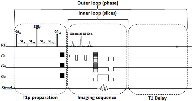
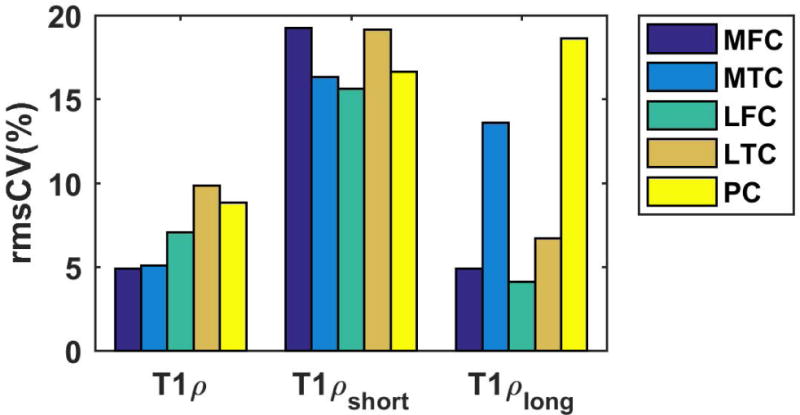
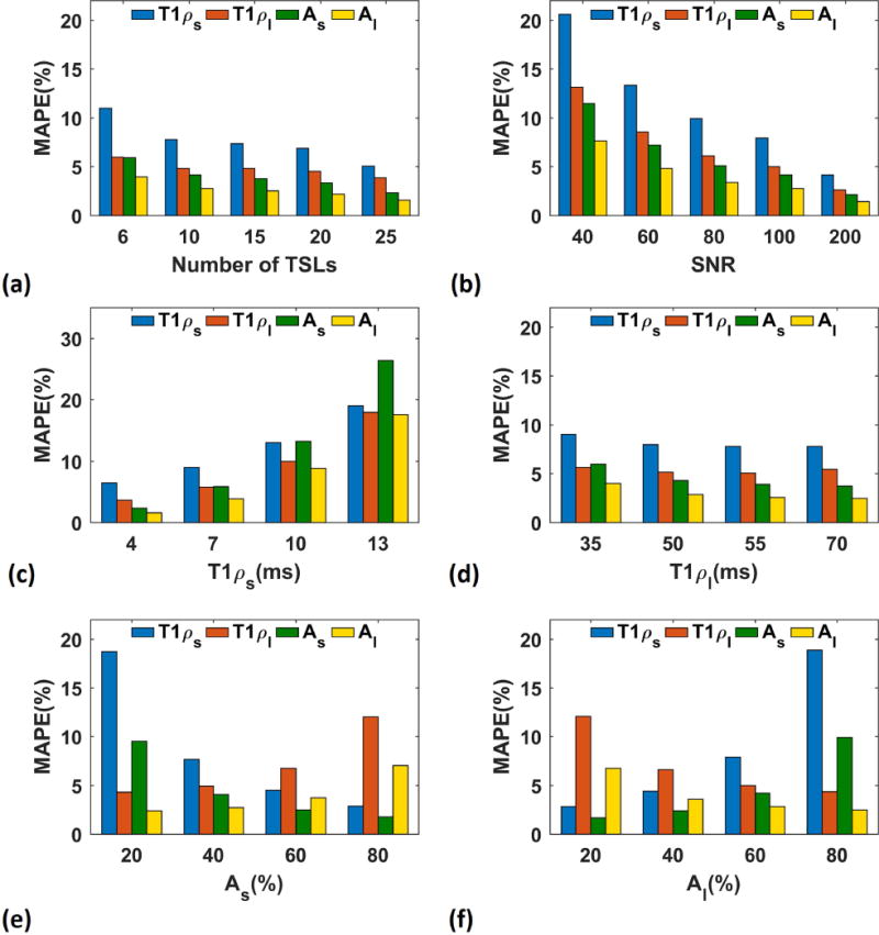
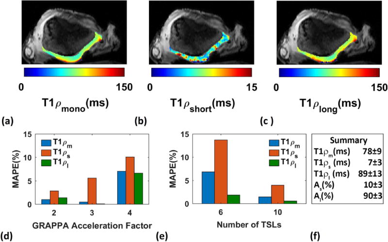
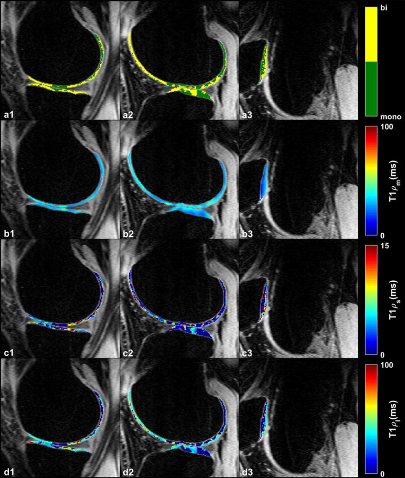
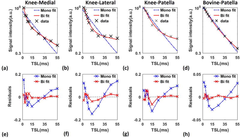
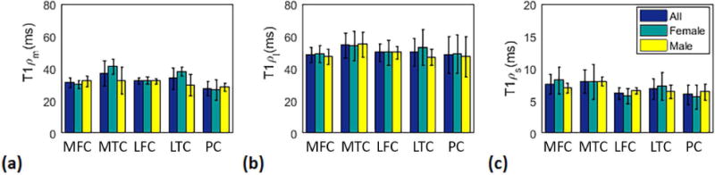
The purpose of this study was to demonstrate the feasibility of biexponential T relaxation mapping of human knee cartilage in vivo. A three-dimensional, customized, turbo-flash sequence was used to acquire T -weighted images from healthy volunteers employing a standard 3-T MRI clinical scanner. A series of T -weighted images was fitted using monoexponential and biexponential models with two- and four-parametric non-linear approaches, respectively. Non-parametric Kruskal-Wallis and Mann-Whitney U-statistical tests were used to evaluate the regional relaxation and gender differences, respectively, with a level of significance of P = 0.05. Biexponential relaxations were detected in the cartilage of all volunteers. The short and long relaxation components of T were estimated to be 6.9 and 51.0 ms, respectively. Similarly, the fractions of short and long T were 37.6% and 62.4%, respectively. The monoexponential relaxation of T was 32.6 ms. The experiments showed good repeatability with a coefficient of variation (CV) of less than 20%. A biexponential relaxation model showed a better fit than a monoexponential model to the T relaxation decay in knee cartilage. Biexponential T components could potentially be used to increase the specificity to detect early osteoarthritis by the measurement of different water compartments and their fractions.

摘要

本研究的目的是证明在体测量人膝关节软骨双指数T弛豫映射的可行性。使用三维定制的快速扰相梯度回波序列,通过标准的3T磁共振成像临床扫描仪从健康志愿者获取T加权图像。分别采用双参数和四参数非线性方法,用单指数和双指数模型拟合一系列T加权图像。采用非参数Kruskal-Wallis检验和Mann-Whitney U统计检验分别评估区域弛豫差异和性别差异,显著性水平为P = 0.05。在所有志愿者的软骨中均检测到双指数弛豫。T的短弛豫成分和长弛豫成分估计分别为6.9 ms和51.0 ms。同样,短T和长T的分数分别为37.6%和62.4%。T的单指数弛豫为32.6 ms。实验显示出良好的重复性,变异系数(CV)小于20%。双指数弛豫模型比单指数模型能更好地拟合膝关节软骨的T弛豫衰减。双指数T成分有可能通过测量不同水相及其分数来提高检测早期骨关节炎的特异性。

https://cdn.ncbi.nlm.nih.gov/pmc/blobs/68e2/5597480/6bdb9aa6c713/nihms888818f8.jpg
https://cdn.ncbi.nlm.nih.gov/pmc/blobs/68e2/5597480/77b9f876bd70/nihms888818f1.jpg
https://cdn.ncbi.nlm.nih.gov/pmc/blobs/68e2/5597480/57b01ea6ff2f/nihms888818f2.jpg
https://cdn.ncbi.nlm.nih.gov/pmc/blobs/68e2/5597480/a3a1afcd9556/nihms888818f3.jpg
https://cdn.ncbi.nlm.nih.gov/pmc/blobs/68e2/5597480/94aa387be8e0/nihms888818f4.jpg
https://cdn.ncbi.nlm.nih.gov/pmc/blobs/68e2/5597480/9267e9c10ffb/nihms888818f5.jpg
https://cdn.ncbi.nlm.nih.gov/pmc/blobs/68e2/5597480/a95ee95866cd/nihms888818f6.jpg
https://cdn.ncbi.nlm.nih.gov/pmc/blobs/68e2/5597480/37029998777d/nihms888818f7.jpg
https://cdn.ncbi.nlm.nih.gov/pmc/blobs/68e2/5597480/6bdb9aa6c713/nihms888818f8.jpg

相似文献

1
Biexponential T relaxation mapping of human knee cartilage in vivo at 3 T.3T下人体膝关节软骨的双指数T弛豫映射的体内研究
NMR Biomed. 2017 Oct;30(10). doi: 10.1002/nbm.3760. Epub 2017 Jun 20.
2
Biexponential T relaxation estimation of human knee cartilage in vivo at 3T.人体膝关节软骨在 3T 下的双指数 T 弛豫估计。
J Magn Reson Imaging. 2018 Mar;47(3):809-819. doi: 10.1002/jmri.25778. Epub 2017 May 31.
3
Biexponential T relaxation mapping of human knee menisci.人体半月板的双指数 T 弛豫映射。
J Magn Reson Imaging. 2019 Sep;50(3):824-835. doi: 10.1002/jmri.26631. Epub 2019 Jan 7.
4
Compressed sensing acceleration of biexponential 3D-T relaxation mapping of knee cartilage.压缩感知加速膝关节软骨双指数 3D-T2 弛豫成像。
Magn Reson Med. 2019 Feb;81(2):863-880. doi: 10.1002/mrm.27416. Epub 2018 Sep 19.
5
Isotropic morphometry and multicomponent T ρ mapping of human knee articular cartilage in vivo at 3T.在 3T 下对人体膝关节关节软骨进行各向同性形态测量和多分量 T ρ 映射。
J Magn Reson Imaging. 2018 Dec;48(6):1707-1716. doi: 10.1002/jmri.26173. Epub 2018 May 2.
6
3D-T prepared zero echo time-based PETRA sequence for in vivo biexponential relaxation mapping of semisolid short-T tissues at 3 T.3T 下用于半固态短 T 组织体内双指数弛豫测绘的 3D-T 准备零回波时间基于 PETRA 的序列。
J Magn Reson Imaging. 2019 Oct;50(4):1207-1218. doi: 10.1002/jmri.26664. Epub 2019 Jan 28.
7
Bi-component T1ρ and T2 Relaxation Mapping of Skeletal Muscle In-Vivo.骨骼肌在体双组份 T1ρ 和 T2 弛豫测绘。
Sci Rep. 2017 Oct 26;7(1):14115. doi: 10.1038/s41598-017-14581-9.
8
Accelerated mono- and biexponential 3D-T1ρ relaxation mapping of knee cartilage using golden angle radial acquisitions and compressed sensing.利用黄金角径向采集和压缩感知技术对膝关节软骨进行加速单指数和双指数3D-T1ρ弛豫映射。
Magn Reson Med. 2020 Apr;83(4):1291-1309. doi: 10.1002/mrm.28019. Epub 2019 Oct 18.
9
3D magnetic resonance fingerprinting for rapid simultaneous T1, T2, and T1ρ volumetric mapping of human articular cartilage at 3 T.3T 下用于人体关节软骨的快速、同步 T1、T2 和 T1ρ 容积成像的三维磁共振指纹成像技术。
NMR Biomed. 2022 Dec;35(12):e4800. doi: 10.1002/nbm.4800. Epub 2022 Jul 30.
10
Rapid mono and biexponential 3D-T mapping of knee cartilage using variational networks.使用变分网络对膝关节软骨进行快速单指数和双指数 3D-T 映射。
Sci Rep. 2020 Nov 5;10(1):19144. doi: 10.1038/s41598-020-76126-x.

引用本文的文献

1
Feasibility of a UTE Stack-of-Spirals Sequence for Biexponential T Mapping of Whole Knee Joint.UTE 螺旋堆叠序列用于全膝关节双指数 T 映射的可行性
NMR Biomed. 2025 Mar;38(3):e70008. doi: 10.1002/nbm.70008.
2
HDNLS: Hybrid Deep-Learning and Non-Linear Least Squares-Based Method for Fast Multi-Component T1ρ Mapping in the Knee Joint.HDNLS:基于混合深度学习和非线性最小二乘法的膝关节快速多成分T1ρ映射方法
Bioengineering (Basel). 2024 Dec 25;12(1):8. doi: 10.3390/bioengineering12010008.
3
Optimized MR pulse sequence for high-resolution brain 3D-T1ρ mapping with weighted spin-lock acquisitions.

本文引用的文献

1
MRI T1ρ and T2 mapping for the assessment of articular cartilage changes in patients with medial knee osteoarthritis after hemicallotasis osteotomy.MRI T1ρ 和 T2 图谱用于评估内侧膝关节骨关节炎患者半骺阻滞术后关节软骨的变化。
Bone Joint Res. 2016 Jul;5(7):294-300. doi: 10.1302/2046-3758.57.BJR-2016-0057.R1.
2
T1ρ and T2 -based characterization of regional variations in intervertebral discs to detect early degenerative changes.基于T1ρ和T2的椎间盘区域差异特征分析以检测早期退变改变。
J Orthop Res. 2016 Aug;34(8):1373-81. doi: 10.1002/jor.23311. Epub 2016 Jun 14.
3
Paired self-compensated spin-lock preparation for improved T1ρ quantification.
用于高分辨率脑部三维T1ρ图谱绘制的加权自旋锁定采集优化磁共振脉冲序列。
Magn Reson Med. 2025 Apr;93(4):1458-1470. doi: 10.1002/mrm.30412. Epub 2024 Dec 22.
4
Ultrafast - NMR for Correlating Different Motional Regimes of Molecules.用于关联分子不同运动状态的超快核磁共振
Anal Chem. 2024 Oct 22;96(42):16534-16542. doi: 10.1021/acs.analchem.4c00513. Epub 2024 Oct 9.
5
Performance of MR learned pulse sequences for 3D bi-exponential, stretched-exponential, and mono-exponential T and T mapping of knee cartilage.MR 学习脉冲序列在膝关节软骨 3D 双指数、拉伸指数和单指数 T 和 T 映射中的性能。
Magn Reson Med. 2025 Feb;93(2):643-656. doi: 10.1002/mrm.30303. Epub 2024 Sep 23.
6
Qualitative and Quantitative MR Imaging of the Cartilaginous Endplate: A Review.软骨终板的定性和定量磁共振成像:综述
J Magn Reson Imaging. 2025 Apr;61(4):1552-1571. doi: 10.1002/jmri.29562. Epub 2024 Aug 20.
7
Novel spin-lock time sampling strategies for improved reproducibility in quantitative T1ρ mapping.新型自旋锁定时间采样策略可提高定量 T1ρ 映射的可重复性。
NMR Biomed. 2024 Dec;37(12):e5244. doi: 10.1002/nbm.5244. Epub 2024 Aug 17.
8
Bi-Exponential 3D UTE-T1ρ Relaxation Mapping of Ex Vivo Human Knee Patellar Tendon at 3T.3T下离体人膝髌腱的双指数3D UTE-T1ρ弛豫映射
Bioengineering (Basel). 2024 Jan 9;11(1):66. doi: 10.3390/bioengineering11010066.
9
Optimizing variable flip angles in magnetization-prepared gradient-echo sequences for efficient 3D-T1ρ mapping.优化磁化准备梯度回波序列中的变量翻转角,以实现高效的 3D-T1ρ 映射。
Magn Reson Med. 2023 Oct;90(4):1465-1483. doi: 10.1002/mrm.29740. Epub 2023 Jun 8.
10
Optimization of spin-lock times for T mapping of human knee cartilage with bi- and stretched-exponential models.双指数和拉伸指数模型用于人体膝关节软骨 T 映射的自旋锁定时间优化。
Sci Rep. 2022 Oct 7;12(1):16829. doi: 10.1038/s41598-022-21269-2.
用于改进 T1ρ 定量的配对自补偿自旋锁定准备方法。
J Magn Reson. 2016 Jul;268:49-57. doi: 10.1016/j.jmr.2016.04.017. Epub 2016 Apr 30.
4
Quantitative assessment of morphology, T, and T of shoulder cartilage using MRI.使用磁共振成像(MRI)对肩部软骨的形态、T1和T2进行定量评估。
Eur Radiol. 2016 Dec;26(12):4656-4663. doi: 10.1007/s00330-016-4322-6. Epub 2016 Mar 18.
5
Capturing fast relaxing spins with SWIFT adiabatic rotating frame spin-lattice relaxation (T1ρ) mapping.利用SWIFT绝热旋转框架自旋晶格弛豫(T1ρ)映射捕获快速弛豫自旋。
NMR Biomed. 2016 Apr;29(4):420-30. doi: 10.1002/nbm.3474. Epub 2016 Jan 26.
6
Compositional MRI techniques for evaluation of cartilage degeneration in osteoarthritis.用于评估骨关节炎中软骨退变的成分MRI技术。
Osteoarthritis Cartilage. 2015 Oct;23(10):1639-53. doi: 10.1016/j.joca.2015.05.026. Epub 2015 Jun 5.
7
Accelerating T1ρ cartilage imaging using compressed sensing with iterative locally adapted support detection and JSENSE.使用带有迭代局部自适应支持检测和JSENSE的压缩感知加速T1ρ软骨成像
Magn Reson Med. 2016 Apr;75(4):1617-29. doi: 10.1002/mrm.25773. Epub 2015 May 22.
8
T₁ρ MRI of human musculoskeletal system.人体肌肉骨骼系统的T₁ρ磁共振成像
J Magn Reson Imaging. 2015 Mar;41(3):586-600. doi: 10.1002/jmri.24677. Epub 2014 Jun 17.
9
Longitudinal evaluation of T1ρ and T2 spatial distribution in osteoarthritic and healthy medial knee cartilage.骨关节炎和健康内侧膝关节软骨中T1ρ和T2空间分布的纵向评估。
Osteoarthritis Cartilage. 2014 Jan;22(1):51-62. doi: 10.1016/j.joca.2013.10.014. Epub 2013 Nov 2.
10
Simultaneous acquisition of T1ρ and T2 quantification in knee cartilage: repeatability and diurnal variation.膝关节软骨中T1ρ与T2定量的同步采集:重复性与日变化
J Magn Reson Imaging. 2014 May;39(5):1287-93. doi: 10.1002/jmri.24253. Epub 2013 Jul 29.