Acuna Andrea, Drakopoulos Michael A, Leng Yue, Goergen Craig J, Calve Sarah
Weldon School of Biomedical Engineering, Purdue University, 206 South Martin Jischke Drive, West Lafayette, IN 47907, USA.
Weldon School of Biomedical Engineering, Purdue University, 206 South Martin Jischke Drive, West Lafayette, IN 47907, USA.
Dev Biol. 2018 Mar 15;435(2):122-129. doi: 10.1016/j.ydbio.2017.12.022. Epub 2018 Jan 17.
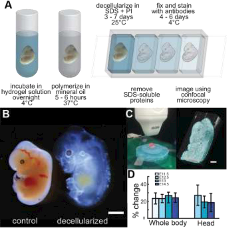
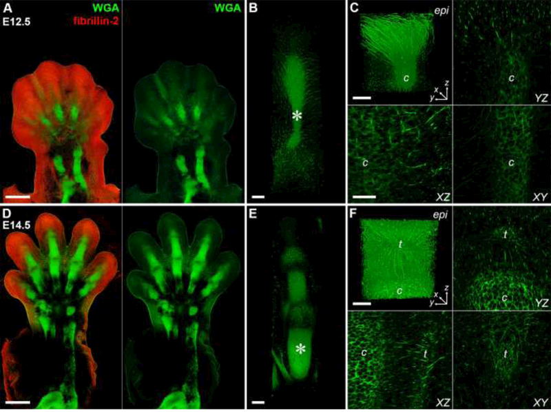
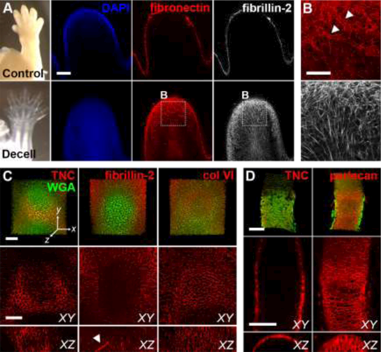
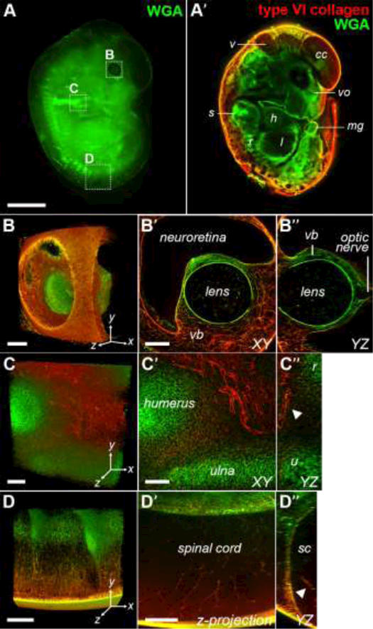
The extracellular matrix (ECM) plays a crucial role in embryogenesis, serving both as a substrate to which cells attach and as an active regulator of cell behavior. However, little is known about the spatiotemporal expression patterns and 3D structure of ECM proteins during embryonic development. The lack of suitable methods to visualize the embryonic ECM is largely responsible for this gap, posing a major technical challenge for biologists and tissue engineers. Here, we describe a method of viewing the 3D organization of the ECM using a polyacrylamide-based hydrogel to provide a 3D framework within developing murine embryos. After removal of soluble proteins using sodium dodecyl sulfate, confocal microscopy was used to visualize the 3D distribution of independent ECM networks in multiple developing tissues, including the forelimb, eye, and spinal cord. Comparative analysis of E12.5 and E14.5 autopods revealed proteoglycan-rich fibrils maintain connections between the epidermis and the underlying tendon and cartilage, indicating a role for the ECM during musculoskeletal assembly and demonstrating that our method can be a powerful tool for defining the spatiotemporal distribution of the ECM during embryogenesis.
细胞外基质(ECM)在胚胎发育中起着至关重要的作用,它既是细胞附着的底物,也是细胞行为的活跃调节因子。然而,关于胚胎发育过程中ECM蛋白的时空表达模式和三维结构,我们知之甚少。缺乏合适的方法来可视化胚胎ECM在很大程度上导致了这一差距,这给生物学家和组织工程师带来了重大技术挑战。在这里,我们描述了一种使用基于聚丙烯酰胺的水凝胶来观察ECM三维组织的方法,以在发育中的小鼠胚胎内提供一个三维框架。使用十二烷基硫酸钠去除可溶性蛋白后,共聚焦显微镜用于可视化多个发育组织中独立ECM网络的三维分布,包括前肢、眼睛和脊髓。对E12.5和E14.5 autopods的比较分析表明,富含蛋白聚糖的纤维维持了表皮与下方肌腱和软骨之间的连接,表明ECM在肌肉骨骼组装过程中发挥了作用,并证明我们的方法可以成为定义胚胎发育过程中ECM时空分布的有力工具。