Porter Charlotte A, Bradley Kevin M, Hippeläinen Eero T, Walker Matthew D, McGowan Daniel R
Radiation Physics and Protection, Churchill Hospital, Oxford University Hospitals NHS Foundation Trust, Oxford, OX3 7LE, UK.
Department of Radiology, Churchill Hospital, Oxford University Hospitals NHS Foundation Trust, Oxford, UK.
EJNMMI Res. 2018 Jan 22;8(1):7. doi: 10.1186/s13550-018-0361-0.
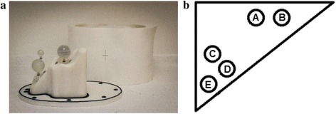
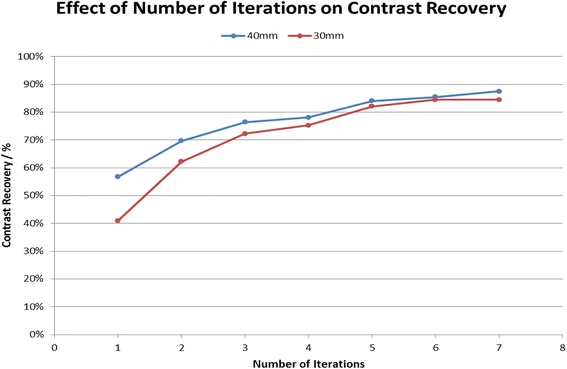
Post-therapy SPECT/CT imaging of Y microspheres delivered to hepatic malignancies is difficult, owing to the continuous, high-energy Bremsstrahlung spectrum emitted by Y. This study aimed to evaluate the utility of a commercially available software package (HybridRecon, Hermes Medical Solutions AB) which incorporates full Monte Carlo collimator modelling. Analysis of image quality was performed on both phantom and clinical images in order to ultimately provide a recommendation of an optimum reconstruction for post-therapy Y microsphere SPECT/CT imaging. A 3D-printed anthropomorphic liver phantom was filled with Y with a sphere-to-background ratio of 4:1 and imaged on a GE Discovery 670 SPECT/CT camera. Datasets were reconstructed using ordered-subsets expectation maximization (OSEM) 1-7 iterations in order to identify the optimal OSEM reconstruction (5 iterations, 15 subsets). Quantitative analysis was subsequently carried out on phantom datasets obtained using four reconstruction algorithms: the default OSEM protocol (2 iterations, 10 subsets) and the optimised OSEM protocol, both with and without full Monte Carlo collimator modelling. The quantitative metrics contrast recovery (CR) and background variability (BV) were calculated. The four algorithms were then used to retrospectively reconstruct 10 selective internal radiation therapy (SIRT) patient datasets which were subsequently blind scored for image quality by a consultant radiologist.
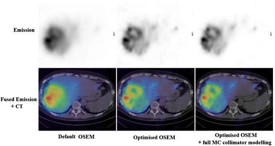
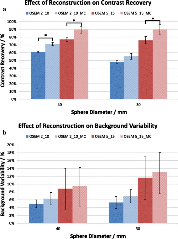
The optimised OSEM reconstruction (5 iterations, 15 subsets with full MC collimator modelling) increased the CR by 42% (p < 0.001) compared to the default OSEM protocol (2 iterations, 10 subsets). The use of full Monte Carlo collimator modelling was shown to further improve CR by 14% (30 mm sphere, CR = 90%, p < 0.05). The consultant radiologist had a significant preference for the optimised OSEM over the default OSEM protocol (p < 0.001), with the optimised OSEM being the favoured reconstruction in every one of the 10 clinical cases presented.
OSEM (5 iterations, 15 subsets) with full Monte Carlo collimator modelling is quantitatively the optimal image reconstruction for post-SIRT Y Bremsstrahlung SPECT/CT imaging. The use of full Monte Carlo collimator modelling for correction of image-degrading effects significantly increases contrast recovery without degrading clinical image quality.
由于钇发射的连续高能轫致辐射谱,对肝恶性肿瘤进行钇微球治疗后的单光子发射计算机断层扫描/计算机断层扫描(SPECT/CT)成像具有挑战性。本研究旨在评估一种商业软件包(HybridRecon,赫耳墨斯医疗解决方案公司)的效用,该软件包纳入了完整的蒙特卡罗准直器建模。为最终推荐钇微球治疗后SPECT/CT成像的最佳重建方法,对体模和临床图像进行了图像质量分析。一个3D打印的拟人化肝脏体模填充了钇,球与背景的比例为4:1,并在GE Discovery 670 SPECT/CT相机上成像。使用有序子集期望最大化(OSEM)算法进行1至7次迭代重建数据集,以确定最佳的OSEM重建(5次迭代,15个子集)。随后,对使用四种重建算法获得的体模数据集进行定量分析:默认的OSEM协议(2次迭代,10个子集)以及优化的OSEM协议,两种协议均有无完整蒙特卡罗准直器建模。计算定量指标对比度恢复(CR)和背景变异性(BV)。然后使用这四种算法对10例选择性内放射治疗(SIRT)患者数据集进行回顾性重建,随后由一位放射科顾问医生对图像质量进行盲法评分。
与默认的OSEM协议(2次迭代,10个子集)相比,优化的OSEM重建(5次迭代,15个子集,采用完整的蒙特卡罗准直器建模)使CR提高了42%(p < 0.001)。结果表明,使用完整的蒙特卡罗准直器建模可使CR进一步提高14%(30毫米球体,CR = 90%,p < 0.05)。放射科顾问医生对优化的OSEM的偏好明显高于默认的OSEM协议(p < 0.001),在呈现的10个临床病例中的每一个病例中,优化的OSEM都是首选的重建方法。
采用完整蒙特卡罗准直器建模的OSEM(5次迭代,15个子集)在定量上是钇轫致辐射SIRT后SPECT/CT成像的最佳图像重建方法。使用完整的蒙特卡罗准直器建模来校正图像退化效应可显著提高对比度恢复,而不会降低临床图像质量。