Cho Geum Joon, Lee Lyn Hwa, Lee Bona, Lee Jaeeun, Ahn Ki-Hoon, Hong Soon-Cheol, Kim Hai-Joong, Oh Min-Jeong
Department of Obstetrics and Gynecology, Korea University College of Medicine, Seoul, Korea.
J & L Women's Clinics, Seoul, Korea.
Obstet Gynecol Sci. 2018 Jan;61(1):71-78. doi: 10.5468/ogs.2018.61.1.71. Epub 2017 Dec 11.
The purpose of this study was to investigate the effects of estradiol on the expression of hypoxia-inducible factor (HIF)-1α and the differentiation of trophoblasts in human first trimester villous explant cultures.
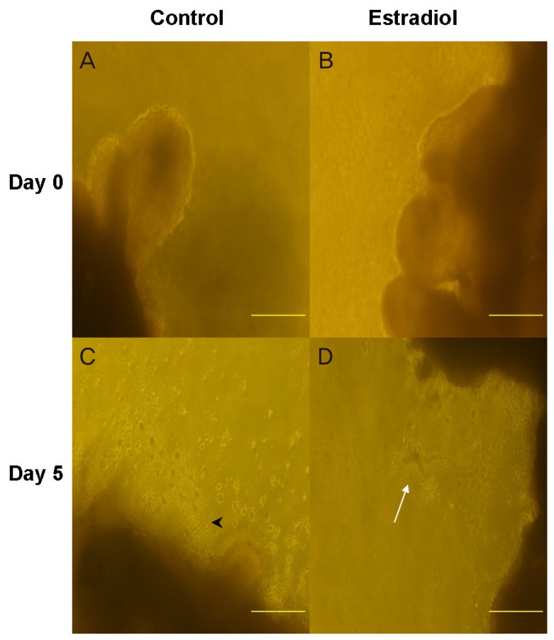
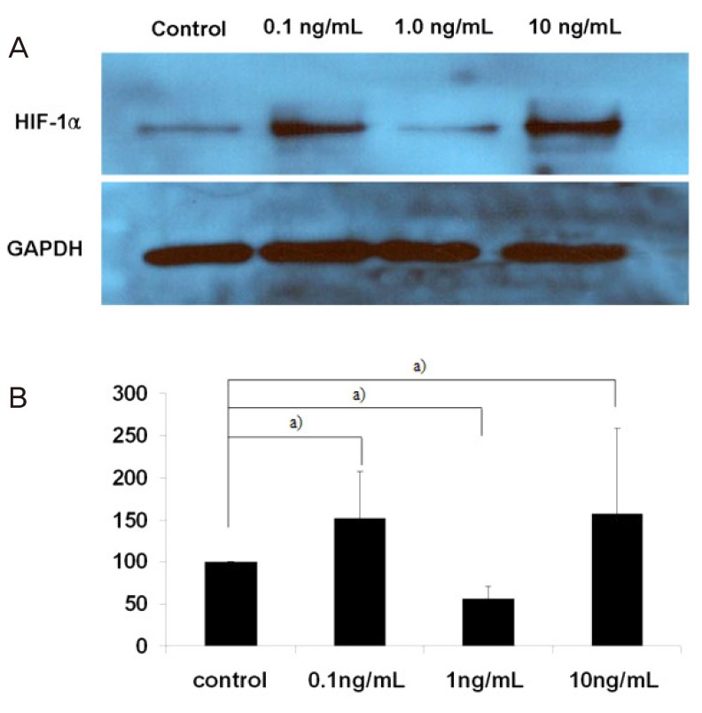
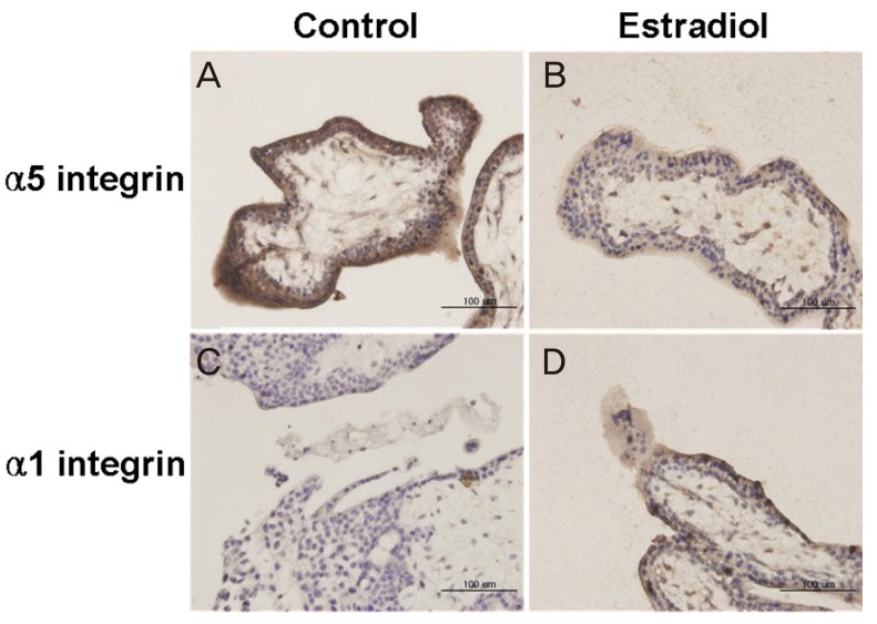
Villous explant cultures were established from first trimester human placentas (6-8 weeks of gestation, n=3). Normal villous tissues were explanted on Matrigel and incubated under 3% O tension for 5 days. To evaluate the effects of estradiol on the villous explant cultures, 1 ng/mL of estradiol was added to the culture medium. The morphological integrities and viabilities of the villous explants were monitored. Immunohistochemistry for α5 and α1 integrin was performed to assess differentiation of extravillous trophoblasts (EVTs). Expression of HIF-1α in villous explant cultures was evaluated by western blotting and densitometry.
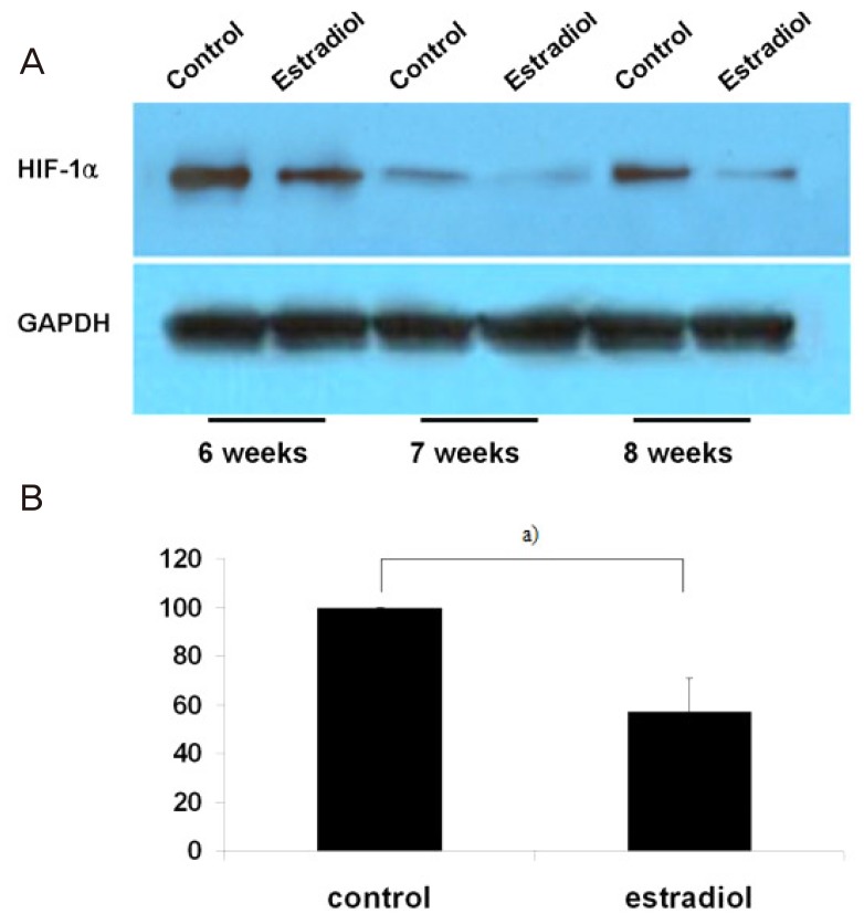
EVTs emerging from first trimester villous explant cultures formed outgrowths of cells from the distal ends and invaded the surrounding Matrigel. Exposure of villous explants to estradiol resulted in the decreased outgrowth of cells from the distal end and decreased expression of α5 integrin. However, estradiol treatment increased the invasion of villous explants into the surrounding Matrigel, concomitant with the increased expression of α1 integrin, indicating differentiation of EVTs into more invasive EVTs. On western blots, the expression of HIF-1α decreased significantly after treatment with estradiol under 3% O tension.
Our findings suggest that estradiol may downregulate expression of HIF-1α in placenta, which in turn promote trophoblast differentiation into invasive phenotype.
本研究旨在探讨雌二醇对人孕早期绒毛外植体培养物中缺氧诱导因子(HIF)-1α表达及滋养层细胞分化的影响。
从人孕早期胎盘(妊娠6 - 8周,n = 3)建立绒毛外植体培养物。将正常绒毛组织接种于基质胶上,在3%氧张力下培养5天。为评估雌二醇对绒毛外植体培养物的影响,向培养基中添加1 ng/mL的雌二醇。监测绒毛外植体的形态完整性和活力。进行α5和α1整合素的免疫组织化学检测以评估绒毛外滋养层细胞(EVT)的分化。通过蛋白质印迹法和光密度测定法评估绒毛外植体培养物中HIF-1α的表达。
孕早期绒毛外植体培养物中出现的EVT从远端形成细胞生长并侵入周围的基质胶。绒毛外植体暴露于雌二醇导致远端细胞生长减少以及α5整合素表达降低。然而,雌二醇处理增加了绒毛外植体向周围基质胶的侵袭,同时伴随着α1整合素表达增加,表明EVT分化为更具侵袭性的EVT。在蛋白质印迹上,在3%氧张力下用雌二醇处理后,HIF-1α的表达显著降低。
我们的研究结果表明,雌二醇可能下调胎盘中HIF-1α的表达,进而促进滋养层细胞分化为侵袭性表型。