Sakamoto Mari, Matsumoto Yoshiko, Mori Sotaro, Ueda Kaori, Inoue Yukako, Kurimoto Takuji, Kanamori Akiyasu, Yamada Yuko, Nakamura Makoto
Department of Surgery, Division of Ophthalmology, Kobe University Graduate School of Medicine, Kobe, Japan.
PLoS One. 2018 Jan 26;13(1):e0191862. doi: 10.1371/journal.pone.0191862. eCollection 2018.
We previously reported that eyes with hypotony maculopathy (HM) after trabeculectomy (TLE) exhibited more reduction of axial length (AL) than those without HM, suggesting that inward collapse of the scleral wall may contribute to the development of HM after TLE. However, we did not evaluate change in choroidal thickness (CT), which could influence AL measures. We compared the magnitude and rate of AL and CT changes in eyes with and without HM by simultaneously measuring these parameters before and after TLE.
We enrolled 77 eyes of 77consecutive patients with glaucoma, who underwent TLE between March 2014 and March 2016. Intraocular pressure (IOP), central corneal thickness, keratometry, AL, and CT were measured pre- and postoperatively, up to 6 months. These biometrics were compared in eyes with and without HM.
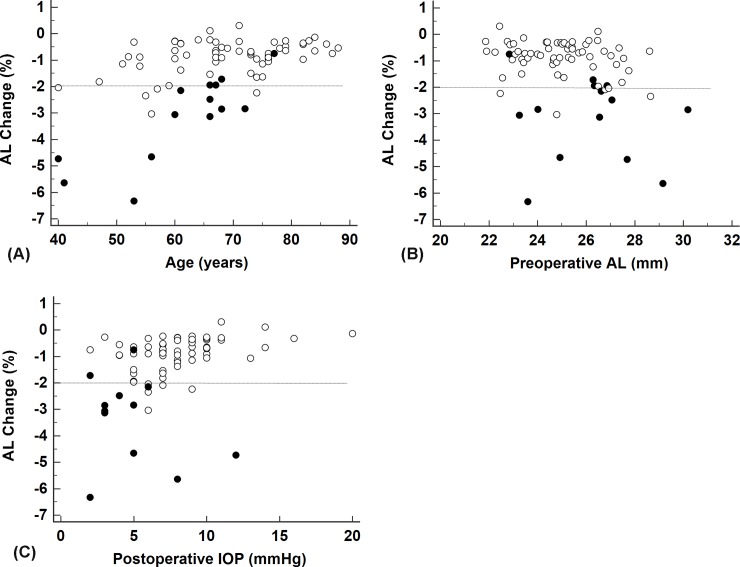
The 14 patients who developed HM were significantly younger than those who did not. The eyes with HM exhibited significantly reduced AL (2.8%) compared to those without HM (0.7%). There was no significant difference in CT change between the two groups. The rate of AL reduction was significantly correlated with age, postoperative IOP, and preoperative AL. Post-adjustment logistic regression analysis revealed that eyes with AL reduction rate ≥ 2% had 11.67 higher risk for developing HM (95% confidence interval, 1.28-106.6; P = 0.03).
AL reduction rates ≥ 2% were significantly associated with HM. Excessive reduction in AL, which was seen in eyes with HM, was not an artificial measure resulting from choroidal thickening but rather reflected reductions in the anterior-posterior diameter of the eyeball. Inward collapse of the scleral wall leads to redundancy of the chorioretinal tissue, contributing to the development of HM after TLE.
我们之前报道过,小梁切除术后发生低眼压性黄斑病变(HM)的眼睛比未发生HM的眼睛轴向长度(AL)减少更多,这表明巩膜壁向内塌陷可能导致小梁切除术后HM的发生。然而,我们没有评估脉络膜厚度(CT)的变化,而脉络膜厚度变化可能会影响AL测量结果。我们通过在小梁切除术前和术后同时测量这些参数,比较了发生和未发生HM的眼睛中AL和CT变化的幅度及速率。
我们纳入了2014年3月至2016年3月期间连续77例接受小梁切除术的青光眼患者的77只眼睛。术前和术后测量眼压(IOP)、中央角膜厚度、角膜曲率、AL和CT,最长随访6个月。对发生和未发生HM的眼睛的这些生物测量指标进行比较。
发生HM的14例患者明显比未发生HM的患者年轻。发生HM的眼睛的AL较未发生HM的眼睛显著降低(2.8%)。两组之间CT变化无显著差异。AL减少速率与年龄、术后IOP和术前AL显著相关。调整后的逻辑回归分析显示,AL减少速率≥2%的眼睛发生HM的风险高11.67倍(95%置信区间,1.28 - 106.6;P = 0.03)。
AL减少速率≥2%与HM显著相关。HM患者眼睛中出现的AL过度减少并非脉络膜增厚导致的人为测量结果,而是反映了眼球前后径的减小。巩膜壁向内塌陷导致脉络膜视网膜组织冗余,促使小梁切除术后HM的发生。