Hebelka Hanna, Miron Andreia, Kasperska Izabela, Brisby Helena, Lagerstrand Kerstin
Department of Radiology, Sahlgrenska University Hospital, Gothenburg, Sweden.
Institute of Clinical Sciences Sahlgrenska Academy, University of Gothenburg, Gothenburg, Sweden.
J Orthop Surg Res. 2018 Jan 30;13(1):18. doi: 10.1186/s13018-018-0727-z.
The function of the endplate (EP) is the most important factor influencing nutritional supply to the avascular intervertebral disc (IVD). It is desired to have a non-invasive method to assess functional EP characteristics in vivo. Assessment of functional EP characteristics is important in order to understand its relation to IVD degeneration, which in turn might deepen the understanding of the pathophysiology behind low back pain (LBP). It was hypothesized that, by comparing quantitative MRI of EPs performed with conventional supine MRI (unloaded MRI) with axial loading during MRI (alMRI), dynamical properties of the EP can be displayed. The aim was therefore to investigate the feasibility of axial loading during MRI (alMRI) to instantaneously induce quantitative EP changes.
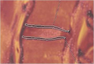
T2 mapping of 55 vertebral EPs (L1-S1) in five LBP patients was performed during conventional supine MRI (unloaded MRI) and subsequent alMRI. With T2 mapping, the cartilaginous EP and bony EP cannot be separated; hence, the visualized EP was termed EP zone (EPZ). Each EPZ was segmented at multiple midsagittal views, generating volumetric regions of interest. EPZs demonstrating signal inhomogeneity and/or adjacent Modic changes (MC) were termed abnormal EPZs. EPZ mean T2 values were compared between unloaded MRI and alMRI, and their relationship with abnormal EPZs was determined.
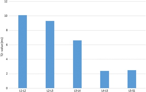
alMRI induced significantly higher (p = 0.01) EPZ mean T2 values compared with unloaded MRI. Significantly higher mean T2 values were seen in inferior EPZs compared with superior EPZs, both with unloaded MRI (35%, p < 0.001) and with alMRI (26%, p = 0.04). Significant difference between unloaded MRI and alMRI was seen in normal (p = 0.02), but not in abnormal EPZs (p = 0.5; n = 12).
alMRI induces changes in human EPZ characteristics in vivo. The T2 value significantly increased in normal EPZs, with lack of such in abnormal EPZs. Combining T2 mapping with alMRI provides a clinical feasible, non-invasive method with potential to reveal biochemical behavioral patterns, thus adding another dimension of the EPZs characteristics compared with information obtained with solely unloaded MRI.
终板(EP)的功能是影响无血管椎间盘(IVD)营养供应的最重要因素。人们期望有一种非侵入性方法来评估体内功能性终板的特征。评估功能性终板特征对于理解其与椎间盘退变的关系很重要,而这反过来可能会加深对腰痛(LBP)背后病理生理学的理解。据推测,通过将常规仰卧位MRI(非负重MRI)与MRI期间轴向负荷(alMRI)下进行的终板定量MRI进行比较,可以显示终板的动态特性。因此,目的是研究MRI期间轴向负荷(alMRI)瞬时诱导终板定量变化的可行性。
对5例腰痛患者的55个椎体终板(L1-S1)在常规仰卧位MRI(非负重MRI)及随后的alMRI期间进行T2 mapping。通过T2 mapping,软骨终板和骨终板无法区分;因此,可视化的终板称为终板区(EPZ)。在多个矢状面视图上对每个终板区进行分割,生成感兴趣的体积区域。表现出信号不均匀和/或相邻Modic改变(MC)的终板区称为异常终板区。比较非负重MRI和alMRI之间终板区的平均T2值,并确定它们与异常终板区的关系。
与非负重MRI相比,alMRI诱导的终板区平均T2值显著更高(p = 0.01)。与上终板区相比,下终板区的平均T2值在非负重MRI(35%,p < 0.001)和alMRI(26%,p = 0.04)时均显著更高。在正常终板区,非负重MRI和alMRI之间存在显著差异(p = 0.02),但在异常终板区无显著差异(p = 0.5;n = 12)。
alMRI可在体内诱导人终板区特征发生变化。正常终板区的T2值显著增加,而异常终板区则无此现象。将T2 mapping与alMRI相结合提供了一种临床可行的非侵入性方法,有可能揭示生化行为模式,从而与仅通过非负重MRI获得的信息相比,增加了终板区特征的另一个维度。