Eladak Soria, Moison Delphine, Guerquin Marie-Justine, Matilionyte Gabriele, Kilcoyne Karen, N'Tumba-Byn Thierry, Messiaen Sébastien, Deceuninck Yoann, Pozzi-Gaudin Stéphanie, Benachi Alexandra, Livera Gabriel, Antignac Jean-Philippe, Mitchell Rod, Rouiller-Fabre Virginie, Habert René
Univ. Paris Diderot, Sorbonne Paris Cité, Laboratory of Development of the Gonads, Unit of Genetic Stability, Stem Cells and Radiation, Fontenay-aux-Roses, France.
CEA, DSV, iRCM, SCSR, LDG, Fontenay-aux-Roses, France.
PLoS One. 2018 Jan 31;13(1):e0191934. doi: 10.1371/journal.pone.0191934. eCollection 2018.
Using an organotypic culture system termed human Fetal Testis Assay (hFeTA) we previously showed that 0.01 μM BPA decreases basal, but not LH-stimulated, testosterone secreted by the first trimester human fetal testis. The present study was conducted to determine the potential for a long-term antiandrogenic effect of BPA using a xenograft model, and also to study the effect of BPA on germ cell development using both the hFETA and xenograft models.
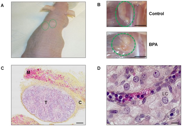
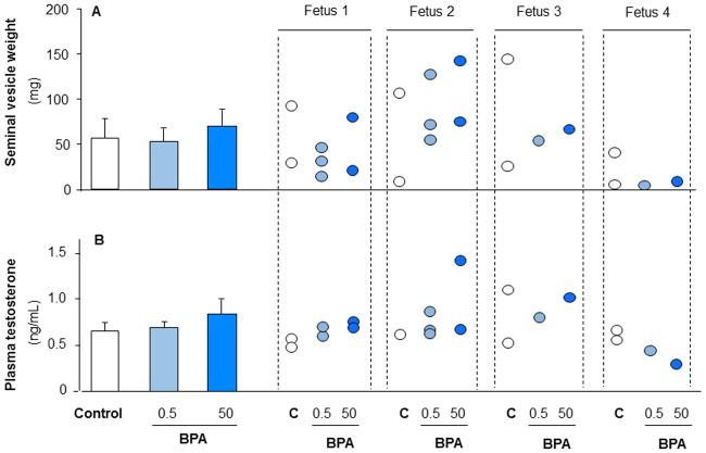
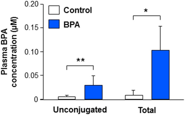
Using the hFeTA system, first trimester testes were cultured for 3 days with 0.01 to 10 μM BPA. For xenografts, adult castrate male nude mice were injected with hCG and grafted with first trimester testes. Host mice received 10 μM BPA (~ 500 μg/kg/day) in their drinking water for 5 weeks. Plasma levels of total and unconjugated BPA were 0.10 μM and 0.038 μM respectively. Mice grafted with second trimester testes received 0.5 and 50 μg/kg/day BPA by oral gavage for 5 weeks.
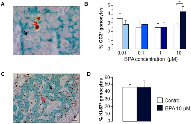
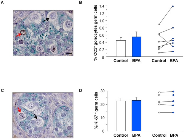
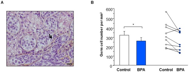
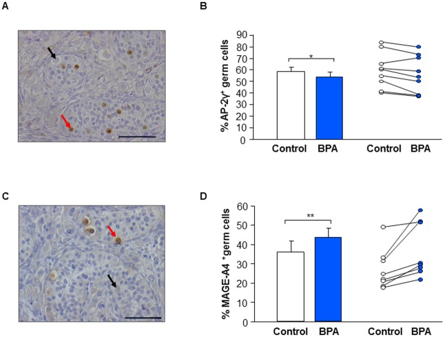
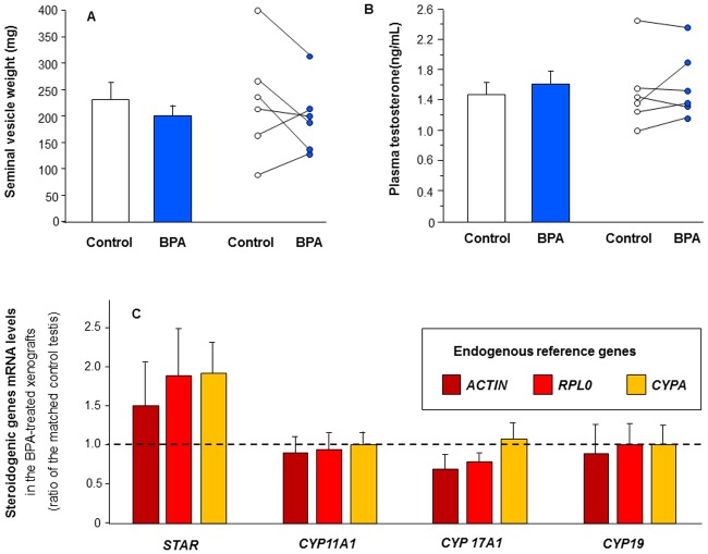
With first trimester human testes, using the hFeTA model, 10 μM BPA increased germ cell apoptosis. In xenografts, germ cell density was also reduced by BPA exposure. Importantly, BPA exposure significantly decreased the percentage of germ cells expressing the pluripotency marker AP-2γ, whilst the percentage of those expressing the pre-spermatogonial marker MAGE-A4 significantly increased. BPA exposure did not affect hCG-stimulated androgen production in first and second trimester xenografts as evaluated by both plasma testosterone level and seminal vesicle weight in host mice.
Exposure to BPA at environmentally relevant concentrations impairs germ cell development in first trimester human fetal testis, whilst gonadotrophin-stimulated testosterone production was unaffected in both first and second trimester testis. Studies using first trimester human fetal testis demonstrate the complementarity of the FeTA and xenograft models for determining the respective short-term and long term effects of environmental exposures.
我们之前使用一种名为人类胎儿睾丸检测(hFeTA)的器官型培养系统表明,0.01μM双酚A(BPA)可降低孕早期人类胎儿睾丸分泌的基础睾酮水平,但不影响促黄体生成素(LH)刺激的睾酮分泌。本研究旨在使用异种移植模型确定BPA长期抗雄激素作用的可能性,并使用hFETA和异种移植模型研究BPA对生殖细胞发育的影响。
使用hFeTA系统,将孕早期睾丸与0.01至10μM BPA培养3天。对于异种移植,成年去势雄性裸鼠注射人绒毛膜促性腺激素(hCG)并移植孕早期睾丸。宿主小鼠在饮用水中接受10μM BPA(约500μg/kg/天),持续5周。血浆中总BPA和未结合BPA的水平分别为0.10μM和0.038μM。移植了孕中期睾丸的小鼠通过口服灌胃接受0.5和50μg/kg/天的BPA,持续5周。
在孕早期人类睾丸中,使用hFeTA模型,10μM BPA增加了生殖细胞凋亡。在异种移植中,BPA暴露也降低了生殖细胞密度。重要的是,BPA暴露显著降低了表达多能性标记物AP-2γ的生殖细胞百分比,而表达精原细胞前体标记物MAGE-A4的生殖细胞百分比显著增加。通过宿主小鼠的血浆睾酮水平和精囊重量评估,BPA暴露不影响孕早期和孕中期异种移植中hCG刺激的雄激素产生。
在与环境相关的浓度下暴露于BPA会损害孕早期人类胎儿睾丸中的生殖细胞发育,而在孕早期和孕中期睾丸中,促性腺激素刺激的睾酮产生均未受影响。使用孕早期人类胎儿睾丸的研究证明了FeTA和异种移植模型在确定环境暴露的短期和长期影响方面的互补性。