Yazdi Hamidreza, Nazarian Ara, Kwon John Y, Hochman Mary G, Pakdaman Reza, Hafezi Poopak, Ghahremani Morteza, Joudi Samad, Ghorbanhoseini Mohammad
Department of Knee Surgery, Firoozgar Hospital, Neuromusculoskeletal Research Center, Iran University of Medical Sciences, District 6, Beh Afarin, Tehran, Iran.
Orthopaedic Surgery, Harvard Medical School - Nazarian Lab, Center for Advanced Orthopaedic Studies, BIDMC, 330 Brookline Ave., RN 115, Boston, MA, 02215, USA.
J Orthop Surg Res. 2018 Jan 31;13(1):21. doi: 10.1186/s13018-017-0710-0.
The anatomical axis of the femur is crucial for determining the correct alignment in corrective osteotomies of the knee, total knee arthroplasty (TKA), and retrograde and antegrade femoral intramedullary nailing (IMN). The aim of this study was to propose the concept of different anatomical axes for the proximal and distal parts of the femur; compare these axes in normally aligned subjects and also to propose the clinical application of these axes.
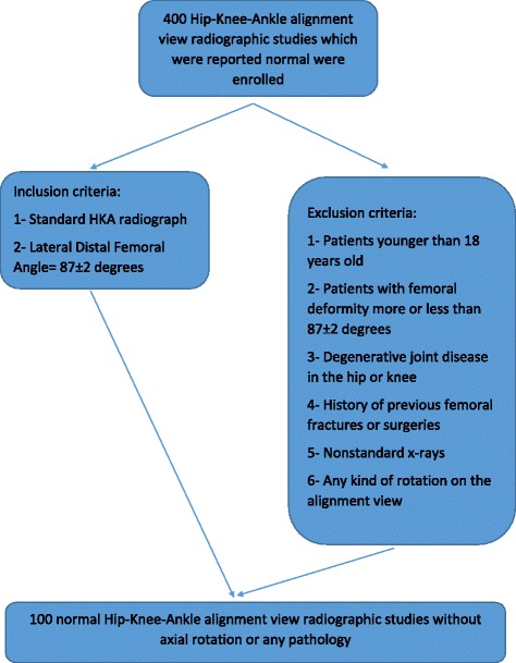
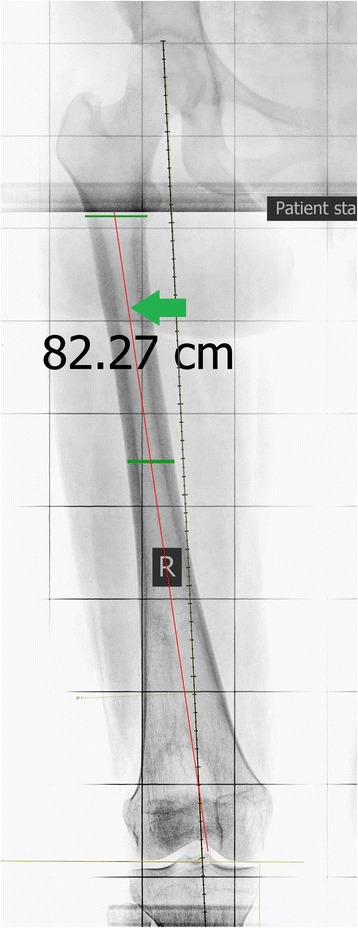
In this cross-sectional study, the horizontal distances between the anatomical axis of the proximal and distal halves of the femur and the center of the intercondylar notch were measured in 100 normally aligned femurs using standard full length alignment view X-rays.
The average age was 34.44 ± 11.14 years. The average distance from the proximal anatomical axis to the center of the intercondylar notch was 6.68 ± 5.23 mm. The proximal anatomical axis of femur passed lateral to the center of the intercondylar notch in 12 cases (12%), medial in 84 cases (84%) and exactly central in 4 cases (4%). The average distance from the distal anatomical axis to the center of the intercondylar notch was 3.63 ± 2.09 mm. The distal anatomical axis of the femur passed medially to the center of the intercondylar notch in 82 cases (82%) and exactly central in 18 cases (18%). There was a significant difference between the anatomical axis of the proximal and distal parts of the femur in reference to the center of intercondylar notch (P value < 0.05), supporting the hypothesis that anatomical axes of the proximal and distal halves of the femur are different in the coronal plane.
While surgeons are aware that the anatomical axis of the distal part of the femur is different than the anatomical axis of the proximal part in patients with femoral deformities, we have shown that these axes are also different in the normally aligned healthy people due to the anatomy of the femur in coronal plane. Also the normal ranges provided here can be used as a reference for the alignment guide entry point in TKA and antegrade and retrograde intramedullary femoral nailing.
股骨的解剖轴对于确定膝关节矫正截骨术、全膝关节置换术(TKA)以及股骨逆行和顺行髓内钉固定术(IMN)中的正确对线至关重要。本研究的目的是提出股骨近端和远端不同解剖轴的概念;比较正常对线受试者的这些轴,并提出这些轴的临床应用。
在这项横断面研究中,使用标准全长对线视图X线片,在100例正常对线的股骨中测量股骨近端和远端两半的解剖轴与髁间切迹中心之间的水平距离。
平均年龄为34.44±11.14岁。从近端解剖轴到髁间切迹中心的平均距离为6.68±5.23毫米。股骨近端解剖轴在12例(12%)中经过髁间切迹中心的外侧,84例(84%)中经过内侧,4例(4%)恰好经过中心。从远端解剖轴到髁间切迹中心的平均距离为3.63±2.09毫米。股骨远端解剖轴在82例(82%)中经过髁间切迹中心的内侧,18例(18%)恰好经过中心。股骨近端和远端部分的解剖轴相对于髁间切迹中心存在显著差异(P值<0.05),支持股骨近端和远端两半的解剖轴在冠状面不同的假设。
虽然外科医生知道在股骨畸形患者中股骨远端部分的解剖轴与近端部分的解剖轴不同,但我们已经表明,由于股骨在冠状面的解剖结构,在正常对线的健康人群中这些轴也不同。此外,这里提供的正常范围可作为TKA以及股骨顺行和逆行髓内钉固定术中对线引导切入点的参考。