Department of Pediatric Orthopedics, The Second Affiliated Hospital of Inner Mongolia Medical University, Hohhot, Inner Mongolia 010030, P.R. China.
Department of Orthopedics and Trauma, The Second Affiliated Hospital of Inner Mongolia Medical University, Hohhot, Inner Mongolia 010030, P.R. China.
Int J Mol Med. 2018 Apr;41(4):2028-2036. doi: 10.3892/ijmm.2018.3412. Epub 2018 Jan 23.
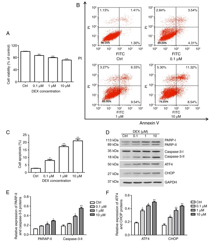
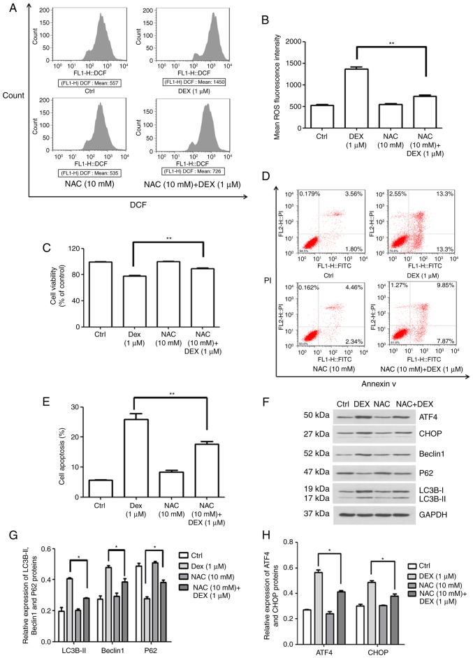
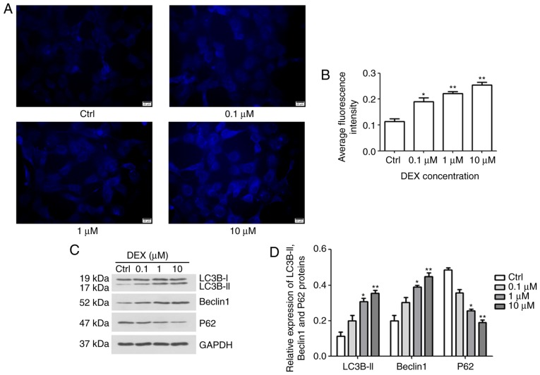
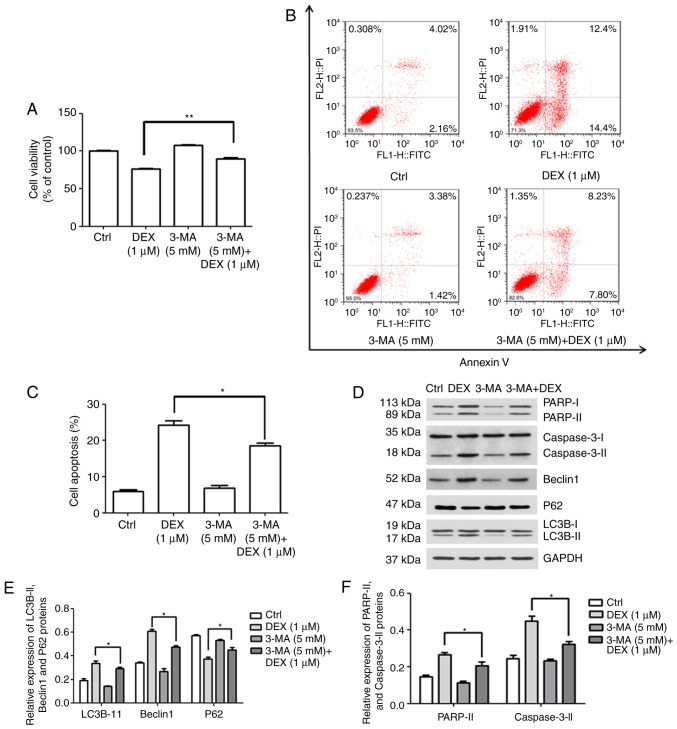
Apoptosis of osteoblasts, triggered by prolonged or excessive use of glucocorticoids (GCs), has been identified as a dominant contributor to the development of osteoporosis and osteonecrosis. However, the molecular mechanisms underlying GC‑induced apoptosis are multifaceted and remain to be fully elucidated. The present study aimed to explore the correlation between dexamethasone (DEX)‑induced reactive oxygen species (ROS), autophagy and apoptosis in MC3T3‑E1 osteoblast‑like cells. Cell viability was assessed using a Cell Counting Kit‑8 assay, and flow cytometry was performed to assess cellular apoptosis, cell cycle and ROS production. Immunofluorescence and western blot analysis were respectively used to detect autophagic vacuoles and the expression of proteins, including cyclin D kinase (CDK)2, poly[ADP ribose] polymerase, caspase‑3, activating transcription factor (ATF)4, CCAAT/enhancer‑binding protein homologous protein (CHOP), Beclin1, microtubule‑associated proteins 1A/1B light chain (LC)3B and P62. It was revealed that DEX not only reduced cell viability, but also promoted apoptosis via the activation of endoplasmic reticulum (ER) stress. In addition, DEX induced cell cycle arrest at G0/G1 phase via inhibition of the expression of CDK2, and the production of ROS was activated. Of note, the DEX‑mediated changes in viability and apoptosis were attenuated in MC3T3‑E1 cells after treatment with 3‑methyladenine, which is an autophagy inhibitor. Treatment with the antioxidant N‑acetylcysteine abolished the effect of DEX on the proliferation, apoptosis, ER stress and autophagy of MC3T3‑E1 cells. In conclusion, the present results indicated that DEX promoted the production of ROS, which enhanced apoptosis through activation of autophagy and ER stress in MC3T3-E1 cells.
成骨细胞的凋亡,是由糖皮质激素(GCs)的长期或过度使用触发的,已被确定为骨质疏松症和骨坏死发展的主要原因。然而,GC 诱导凋亡的分子机制是多方面的,仍有待充分阐明。本研究旨在探讨地塞米松(DEX)诱导 MC3T3-E1 成骨样细胞中活性氧(ROS)、自噬和凋亡之间的相关性。使用细胞计数试剂盒-8 测定细胞活力,通过流式细胞术评估细胞凋亡、细胞周期和 ROS 产生。免疫荧光和 Western blot 分析分别用于检测自噬空泡和包括周期蛋白 D 激酶(CDK)2、多聚(ADP 核糖)聚合酶、半胱天冬酶-3、激活转录因子(ATF)4、CCAAT/增强子结合蛋白同源蛋白(CHOP)、Beclin1、微管相关蛋白 1A/1B 轻链(LC)3B 和 P62 的蛋白表达。结果表明,DEX 不仅降低细胞活力,而且通过激活内质网(ER)应激促进细胞凋亡。此外,DEX 通过抑制 CDK2 的表达诱导细胞周期停滞在 G0/G1 期,并且 ROS 的产生被激活。值得注意的是,在用自噬抑制剂 3-甲基腺嘌呤处理 MC3T3-E1 细胞后,DEX 介导的细胞活力和凋亡变化减弱。用抗氧化剂 N-乙酰半胱氨酸处理可消除 DEX 对 MC3T3-E1 细胞增殖、凋亡、ER 应激和自噬的影响。综上所述,本研究结果表明,DEX 促进了 ROS 的产生,通过激活自噬和 ER 应激促进了 MC3T3-E1 细胞的凋亡。