Macey Paul M, Kheirandish-Gozal Leila, Prasad Janani P, Ma Richard A, Kumar Rajesh, Philby Mona F, Gozal David
School of Nursing, University of California, Los Angeles, CA, United States.
Brain Research Institute, University of California, Los Angeles, CA, United States.
Front Neurol. 2018 Jan 22;9:4. doi: 10.3389/fneur.2018.00004. eCollection 2018.
Obstructive sleep apnea (OSA) affects 2-5% of all children and is associated with cognitive and behavioral deficits, resulting in poor school performance. These psychological deficits may arise from brain injury, as seen in preliminary findings of lower gray matter volume among pediatric OSA patients. However, the psychological deficits in OSA are closely related to functions in the cortex, and such brain areas have not been specifically assessed. The objective was to determine whether cortical thickness, a marker of possible brain injury, is altered in children with OSA.
We examined regional brain cortical thicknesses using high-resolution T1-weighted magnetic resonance images in 16 pediatric OSA patients (8 males; mean age ± SD = 8.4 ± 1.2 years; mean apnea/hypopnea index ± SD = 11 ± 6 events/h) and 138 controls (8.3 ± 1.1 years; 62 male; 138 subjects from the NIH Pediatric MRI database) to identify cortical thickness differences in pediatric OSA subjects.
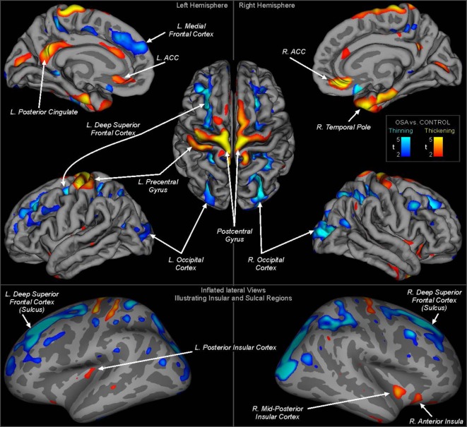
Cortical thinning occurred in multiple regions including the superior frontal, ventral medial prefrontal, and superior parietal cortices. The left side showed greater thinning in the superior frontal cortex. Cortical thickening was observed in bilateral precentral gyrus, mid-to-posterior insular cortices, and left central gyrus, as well as right anterior insula cortex.
Changes in cortical thickness are present in children with OSA and likely indicate disruption to neural developmental processes, including maturational patterns of cortical volume increases and synaptic pruning. Regions with thicker cortices may reflect inflammation or astrocyte activation. Both the thinning and thickening associated with OSA in children may contribute to the cognitive and behavioral dysfunction frequently found in the condition.
阻塞性睡眠呼吸暂停(OSA)影响着2%至5%的儿童,并与认知和行为缺陷相关,导致学业成绩不佳。这些心理缺陷可能源于脑损伤,正如儿科OSA患者灰质体积较低的初步研究结果所示。然而,OSA中的心理缺陷与皮质功能密切相关,而这些脑区尚未得到具体评估。目的是确定OSA儿童是否存在皮质厚度改变,皮质厚度是可能的脑损伤标志物。
我们使用高分辨率T1加权磁共振图像检查了16名儿科OSA患者(8名男性;平均年龄±标准差=8.4±1.2岁;平均呼吸暂停/低通气指数±标准差=11±6次/小时)和138名对照者(8.3±1.1岁;62名男性;来自美国国立卫生研究院儿科MRI数据库的138名受试者)的脑区皮质厚度,以确定儿科OSA受试者的皮质厚度差异。
多个区域出现皮质变薄,包括额上回、腹内侧前额叶和顶上叶皮质。左侧额上回皮质变薄更明显。双侧中央前回、岛叶皮质中后部、左侧中央回以及右侧岛叶前部皮质观察到皮质增厚。
OSA儿童存在皮质厚度变化,这可能表明神经发育过程受到干扰,包括皮质体积增加和突触修剪的成熟模式。皮质较厚的区域可能反映炎症或星形胶质细胞激活。儿童OSA相关的皮质变薄和增厚都可能导致该疾病中常见的认知和行为功能障碍。