Magdaleno Fernando, Ge Xiaodong, Fey Holger, Lu Yongke, Gaskell Harriet, Blajszczak Chuck C, Aloman Costica, Fiel M Isabel, Nieto Natalia
Department of Pathology University of Illinois at Chicago Chicago IL.
Division of Liver Diseases, Department of Medicine Icahn School of Medicine at Mount Sinai New York NY.
Hepatol Commun. 2017 Nov 12;2(1):84-98. doi: 10.1002/hep4.1116. eCollection 2018 Jan.
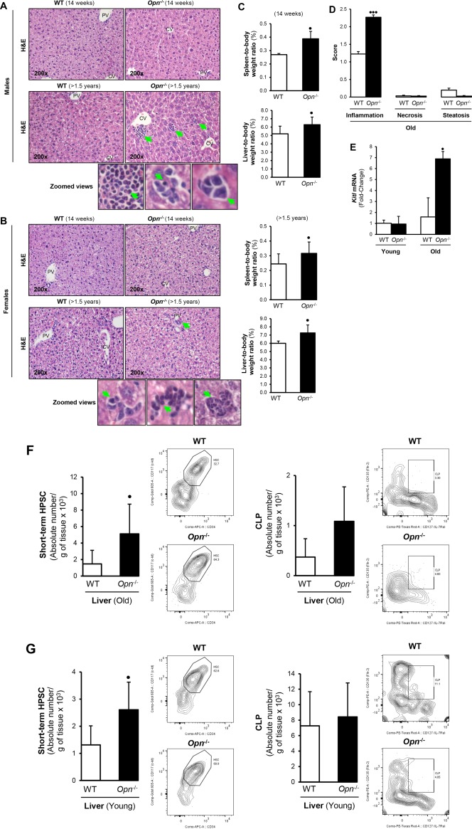
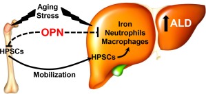
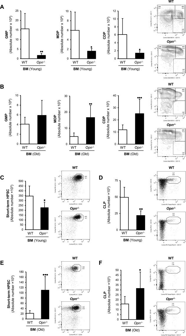
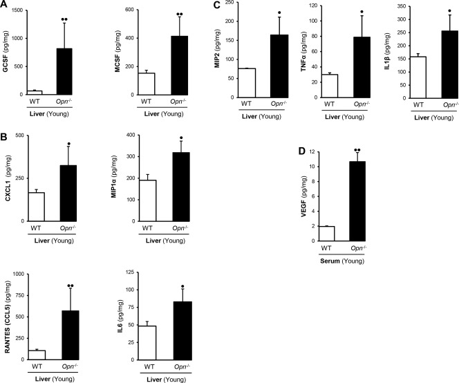
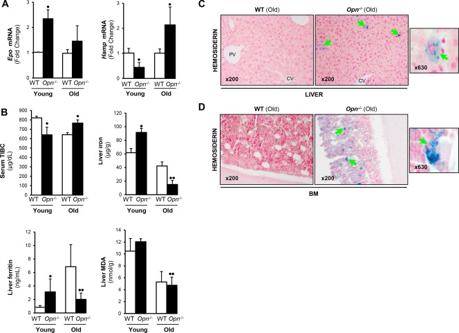
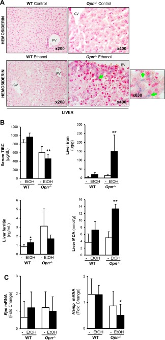
The aim of this study was to investigate the role of osteopontin (OPN) in hematopoietic stem cell (HPSC) mobilization to the liver and its contribution to alcoholic liver disease (ALD). We analyzed young (14-16 weeks) and old (>1.5 years) wild-type (WT) littermates and global knockout ( ) mice for HPSC mobilization to the liver. In addition, WT and mice were chronically fed the Lieber-DeCarli diet for 7 weeks. Bone marrow (BM), blood, spleen, and liver were analyzed by flow cytometry for HPSC progenitors and polymorphonuclear neutrophils (PMNs). Chemokines, growth factors, and cytokines were measured in serum and liver. Prussian blue staining for iron deposits and naphthol AS-D chloroacetate esterase staining for PMNs were performed on liver sections. Hematopoietic progenitors were lower in liver and BM of young compared to old mice. Granulocyte colony-stimulating factor and macrophage colony-stimulating factor were increased in mice, suggesting potential migration of HPSCs from the BM to the liver. Furthermore, ethanol-fed mice showed significant hepatic PMN infiltration and hemosiderin compared to WT mice. As a result, ethanol feeding caused greater liver injury in compared to WT mice. deletion promotes HPSC mobilization, PMN infiltration, and iron deposits in the liver and thereby enhances the severity of ALD. The age-associated contribution of OPN to HPSC mobilization to the liver, the prevalence of PMNs, and accumulation of hepatic iron, which potentiates oxidant stress, reveal novel signaling mechanisms that could be targeted for therapeutic benefit in patients with ALD. ( 2018;2:84-98).
本研究的目的是探讨骨桥蛋白(OPN)在造血干细胞(HPSC)向肝脏动员中的作用及其对酒精性肝病(ALD)的影响。我们分析了年轻(14 - 16周)和年老(>1.5岁)的野生型(WT)同窝小鼠以及全局敲除( )小鼠的HPSC向肝脏的动员情况。此外,对WT和 小鼠长期喂食Lieber - DeCarli饮食7周。通过流式细胞术分析骨髓(BM)、血液、脾脏和肝脏中的HPSC祖细胞和多形核中性粒细胞(PMN)。检测血清和肝脏中的趋化因子、生长因子和细胞因子。对肝脏切片进行普鲁士蓝染色以检测铁沉积,进行萘酚AS - D氯乙酸酯酶染色以检测PMN。与年老的 小鼠相比,年轻小鼠肝脏和BM中的造血祖细胞较少。 小鼠中的粒细胞集落刺激因子和巨噬细胞集落刺激因子增加,提示HPSCs可能从BM迁移至肝脏。此外,与WT小鼠相比,乙醇喂养的 小鼠肝脏显示出显著的PMN浸润和含铁血黄素。结果,与WT小鼠相比,乙醇喂养导致 小鼠肝脏损伤更严重。 缺失促进HPSC动员、PMN浸润和肝脏铁沉积,从而加重ALD的严重程度。OPN对HPSC向肝脏动员的年龄相关影响、PMN的患病率以及肝脏铁的积累(这会增强氧化应激)揭示了新的信号传导机制,这些机制可能成为ALD患者治疗获益的靶点。( 2018;2:84 - 98)