Liu Xiaoyu, Quan Ning
College of Medicine, Institute for Behavioral Medicine Research, The Ohio State University, Columbus, OH, United States.
Division of Biosciences, College of Dentistry, The Ohio State University, Columbus, OH, United States.
Front Neurol. 2018 Jan 23;9:8. doi: 10.3389/fneur.2018.00008. eCollection 2018.
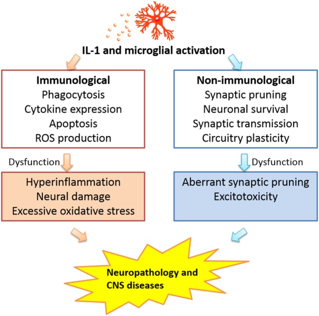
Activation of microglia and expression of the inflammatory cytokine interleukin-1 (IL-1) in the CNS have become almost synonymous with neuroinflammation. In numerous studies, increased CNS IL-1 expression and altered microglial morphology have been used as hallmarks of CNS inflammation. A central concept of how CNS IL-1 and microglia influence functions of the nervous system was derived from the notion initially generated in the peripheral immune system: IL-1 stimulates monocyte/macrophage (the peripheral counterparts of microglia) to amplify inflammation. It is increasingly clear, however, CNS IL-1 acts on other targets in the CNS and microglia participates in many neural functions that are not related to immunological activities. Further, CNS exhibits immunological privilege (although not as absolute as previously thought), rendering amplification of inflammation within CNS under stringent control. This review will analyze current literature to evaluate the contribution of immunological and non-immunological aspects of microglia/IL-1 interaction in the CNS to gain insights for how these aspects might affect health and disease in the nervous tissue.
在中枢神经系统(CNS)中,小胶质细胞的激活以及炎性细胞因子白细胞介素 -1(IL-1)的表达几乎已成为神经炎症的同义词。在众多研究中,中枢神经系统中IL-1表达的增加和小胶质细胞形态的改变已被用作中枢神经系统炎症的标志。关于中枢神经系统IL-1和小胶质细胞如何影响神经系统功能的核心概念最初源于外周免疫系统中产生的观点:IL-1刺激单核细胞/巨噬细胞(小胶质细胞在外周的对应物)以放大炎症。然而,越来越清楚的是,中枢神经系统IL-1作用于中枢神经系统中的其他靶点,并且小胶质细胞参与许多与免疫活动无关的神经功能。此外,中枢神经系统表现出免疫特权(尽管不像以前认为的那样绝对),使得中枢神经系统内炎症的放大受到严格控制。本综述将分析当前文献,以评估中枢神经系统中小胶质细胞/IL-1相互作用的免疫和非免疫方面的作用,从而深入了解这些方面如何影响神经组织的健康和疾病。