• 文献检索
  • 文档翻译
  • 深度研究
  • 学术资讯
  • Suppr Zotero 插件Zotero 插件
  • 邀请有礼
  • 套餐&价格
  • 历史记录
应用&插件
Suppr Zotero 插件Zotero 插件浏览器插件Mac 客户端Windows 客户端微信小程序
定价
高级版会员购买积分包购买API积分包
服务
文献检索文档翻译深度研究API 文档MCP 服务
关于我们
关于 Suppr公司介绍联系我们用户协议隐私条款
关注我们

Suppr 超能文献

核心技术专利:CN118964589B侵权必究
粤ICP备2023148730 号-1Suppr @ 2026

文献检索

告别复杂PubMed语法,用中文像聊天一样搜索,搜遍4000万医学文献。AI智能推荐,让科研检索更轻松。

立即免费搜索

文件翻译

保留排版,准确专业,支持PDF/Word/PPT等文件格式,支持 12+语言互译。

免费翻译文档

深度研究

AI帮你快速写综述,25分钟生成高质量综述,智能提取关键信息,辅助科研写作。

立即免费体验

三维涡轮场回波磁共振成像序列在小儿先天性心脏病合并气管支气管异常中的诊断准确性

Diagnostic Accuracy of Three-dimensional Turbo Field Echo Magnetic Resonance Imaging Sequence in Pediatric Tracheobronchial Anomalies with Congenital Heart Disease.

作者信息

Hou QiaoRu, Gao Wei, Zhong YuMin, Sun AiMin, Wang Qian, Hu LiWei, Wang JingLei

机构信息

Diagnostic imaging Center of Shanghai Children's Medical Center affiliated with Shanghai Jiao Tong University Medical School, Shanghai, China.

Department of Pediatric Cardiology of Shanghai Children's Medical Center affiliated with Shanghai Jiao Tong University Medical School, Shanghai, China.

出版信息

Sci Rep. 2018 Feb 7;8(1):2529. doi: 10.1038/s41598-018-20892-2.

DOI:10.1038/s41598-018-20892-2
PMID:29416073
原文链接:https://pmc.ncbi.nlm.nih.gov/articles/PMC5803237/
Abstract

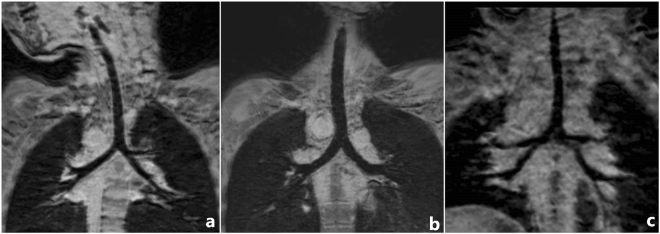
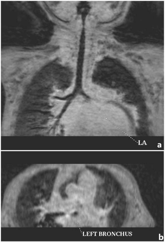
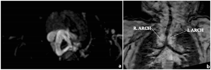
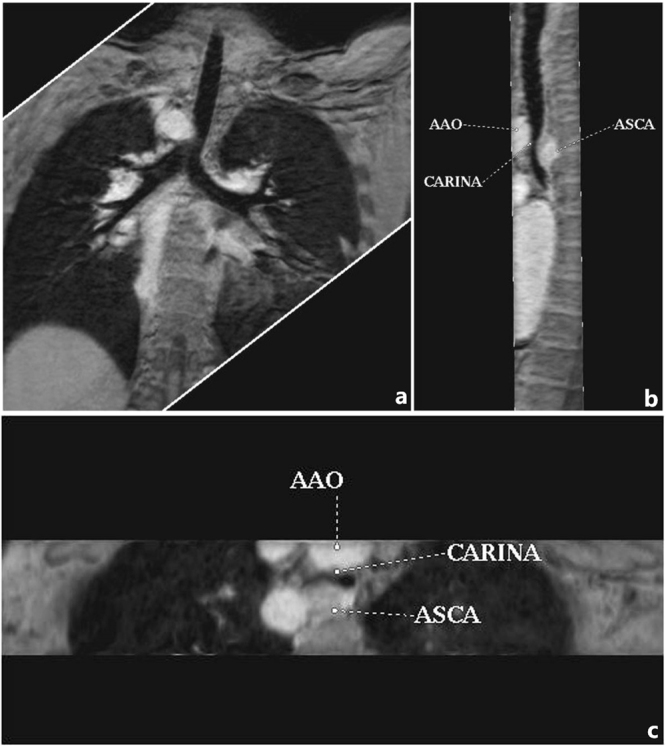
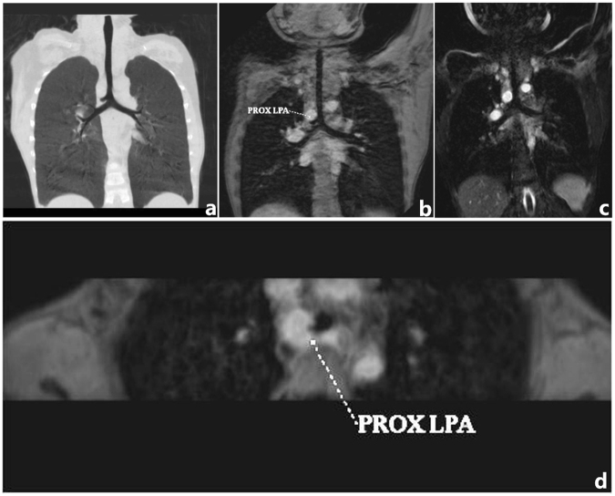
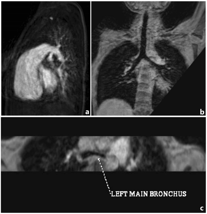
Tracheobronchial anomalies are common in congenital heart disease (CHD), including tracheobronchial stenosis, tracheal bronchus, cardiac bronchus, and bronchial isomerism, which can cause varying degrees of respiratory illness. It is necessary to assess tracheobronchial anomalies and make a preoperative airway evaluation. Multi-slice computed tomography (MSCT) and cardiac magnetic resonance imaging (MRI) are the most effective noninvasive modalities for the diagnosis of CHD and the associated tracheobronchial anomalies. However, MSCT remains an ionizing procedure despite using low dose protocols. The aim of this study was to evaluate diagnostic accuracy of tracheobronchial anomalies in patients with CHD using three-dimensional turbo field echo(3D-TFE) magnetic resonance imaging sequence for preoperative airway evaluation. The results indicated that 3D-TFE provided better image quality as compared to that of 3D-balanced turbo field echo (3D-bTFE), and it can clearly demonstrated the tracheobronchial tree and tracheobronchial anomalies in CHD. This study confirms the clinical value of 3D-TFE in diagnosing tracheobronchial anomalies and supply helpful tracheobronchial information for preoperative strategies and postoperative follow-up.

摘要

气管支气管畸形在先天性心脏病(CHD)中很常见,包括气管支气管狭窄、气管支气管、心脏支气管和支气管异构,可导致不同程度的呼吸系统疾病。评估气管支气管畸形并进行术前气道评估很有必要。多层螺旋计算机断层扫描(MSCT)和心脏磁共振成像(MRI)是诊断CHD及相关气管支气管畸形最有效的无创检查方法。然而,尽管使用了低剂量方案,MSCT仍是一种电离程序。本研究的目的是使用三维快速场回波(3D-TFE)磁共振成像序列评估CHD患者气管支气管畸形的诊断准确性,用于术前气道评估。结果表明,与三维平衡快速场回波(3D-bTFE)相比,3D-TFE提供了更好的图像质量,并且它可以清晰地显示CHD中的气管支气管树和气管支气管畸形。本研究证实了3D-TFE在诊断气管支气管畸形方面的临床价值,并为术前策略和术后随访提供了有用的气管支气管信息。

https://cdn.ncbi.nlm.nih.gov/pmc/blobs/8726/5803237/160716fd9725/41598_2018_20892_Fig6_HTML.jpg
https://cdn.ncbi.nlm.nih.gov/pmc/blobs/8726/5803237/7e7441311bdb/41598_2018_20892_Fig1_HTML.jpg
https://cdn.ncbi.nlm.nih.gov/pmc/blobs/8726/5803237/b109a183fed3/41598_2018_20892_Fig2_HTML.jpg
https://cdn.ncbi.nlm.nih.gov/pmc/blobs/8726/5803237/0ae1f9893d78/41598_2018_20892_Fig3_HTML.jpg
https://cdn.ncbi.nlm.nih.gov/pmc/blobs/8726/5803237/019b82f3045c/41598_2018_20892_Fig4_HTML.jpg
https://cdn.ncbi.nlm.nih.gov/pmc/blobs/8726/5803237/dcf01b4340d1/41598_2018_20892_Fig5_HTML.jpg
https://cdn.ncbi.nlm.nih.gov/pmc/blobs/8726/5803237/160716fd9725/41598_2018_20892_Fig6_HTML.jpg

相似文献

1
Diagnostic Accuracy of Three-dimensional Turbo Field Echo Magnetic Resonance Imaging Sequence in Pediatric Tracheobronchial Anomalies with Congenital Heart Disease.三维涡轮场回波磁共振成像序列在小儿先天性心脏病合并气管支气管异常中的诊断准确性
Sci Rep. 2018 Feb 7;8(1):2529. doi: 10.1038/s41598-018-20892-2.
2
The association of congenital tracheobronchial stenosis and cardiovascular anomalies.先天性气管支气管狭窄与心血管异常的关联。
Int J Pediatr Otorhinolaryngol. 2016 Apr;83:1-6. doi: 10.1016/j.ijporl.2016.01.024. Epub 2016 Jan 27.
3
Tracheobronchial anomalies and stenoses: detection with low-dose multidetector CT with virtual tracheobronchoscopy--comparison with flexible tracheobronchoscopy.气管支气管异常与狭窄:低剂量多层螺旋CT联合虚拟气管支气管镜检查的检测——与可弯曲气管支气管镜检查的比较
Radiology. 2007 Feb;242(2):542-9. doi: 10.1148/radiol.2422060153.
4
Usefulness of three-dimensional electron beam computed tomography for evaluating tracheobronchial anomalies in children with congenital heart disease.三维电子束计算机断层扫描在评估先天性心脏病患儿气管支气管异常中的应用价值。
Am J Cardiol. 2003 Aug 15;92(4):483-6. doi: 10.1016/s0002-9149(03)00676-3.
5
Multislice Computed Tomography Assessment of Tracheobronchial Patterns in Partial Anomalous Left Pulmonary Artery.多层螺旋计算机断层扫描对部分性左肺动脉异常时气管支气管形态的评估
J Comput Assist Tomogr. 2017 Nov/Dec;41(6):983-989. doi: 10.1097/RCT.0000000000000623.
6
Tracheobronchial anomalies in syndromic craniosynostosis with 3-dimensional CT image and bronchoscopy.三维CT图像和支气管镜检查在综合征性颅缝早闭中的气管支气管异常
J Craniofac Surg. 2011 Sep;22(5):1579-83. doi: 10.1097/SCS.0b013e31822e5d15.
7
Morphologic Analysis of Congenital Heart Disease With Anomalous Tracheobronchial Arborization.先天性心脏病伴异常气管支气管树形态学分析。
Ann Thorac Surg. 2020 Oct;110(4):1387-1395. doi: 10.1016/j.athoracsur.2020.01.027. Epub 2020 Feb 27.
8
Combined Surgical and Endoscopic Approach for Ring-Sling Complex.联合手术与内镜治疗吊带复合体的方法
Thorac Cardiovasc Surg. 2020 Jan;68(1):51-58. doi: 10.1055/s-0039-1678670. Epub 2019 Feb 11.
9
Evaluation of tracheobronchial anomalies in children using low-dose multidetector CT: report of a 13-year-old boy with a tracheal bronchus and recurrent pulmonary infections.使用低剂量多层螺旋CT评估儿童气管支气管异常:一名患有气管支气管和反复肺部感染的13岁男孩的病例报告
Pediatr Pulmonol. 2004 Aug;38(2):168-73. doi: 10.1002/ppul.20077.
10
Tracheobronchial abnormalities on computed tomography angiography in children with congenital heart disease.先天性心脏病患儿 CT 血管造影的气管支气管异常。
Pediatr Pulmonol. 2024 May;59(5):1438-1448. doi: 10.1002/ppul.26934. Epub 2024 Feb 20.

引用本文的文献

1
Delayed three-dimensional inversion recovery-prepared fast low-angle shot for airway imaging in children: More than myocardial fibrosis assessment.用于儿童气道成像的延迟三维反转恢复准备快速低角度激发序列:不仅仅用于心肌纤维化评估。
J Cardiovasc Magn Reson. 2024;26(2):101110. doi: 10.1016/j.jocmr.2024.101110. Epub 2024 Oct 23.
2
Altered spontaneous brain activity during dobutamine challenge in healthy young adults: A resting-state functional magnetic resonance imaging study.健康年轻成年人在多巴酚丁胺激发试验期间自发脑活动的改变:一项静息态功能磁共振成像研究。
Front Neurosci. 2023 Jan 6;16:1033569. doi: 10.3389/fnins.2022.1033569. eCollection 2022.
3

本文引用的文献

1
Magnetic resonance imaging in children: common problems and possible solutions for lung and airways imaging.儿童磁共振成像:肺部和气道成像的常见问题及可能的解决方案
Pediatr Radiol. 2015 Dec;45(13):1901-15. doi: 10.1007/s00247-015-3420-y. Epub 2015 Sep 5.
2
A new morphologic classification of congenital tracheobronchial stenosis.先天性气管支气管狭窄的新形态学分类。
Ann Thorac Surg. 2012 Mar;93(3):958-61. doi: 10.1016/j.athoracsur.2011.12.019.
3
Newer CT applications and their alternatives: what is appropriate in children?新型 CT 应用及其替代方案:哪些适用于儿童?
Intelligent Algorithm-Based Multislice Spiral Computed Tomography to Diagnose Coronary Heart Disease.
基于智能算法的多层螺旋 CT 诊断冠心病。
Comput Math Methods Med. 2022 Jan 13;2022:4900803. doi: 10.1155/2022/4900803. eCollection 2022.
4
Tracheal Stenosis and Congenital Heart Disease in Trisomy 21.21三体综合征中的气管狭窄与先天性心脏病
Children (Basel). 2019 Sep 4;6(9):98. doi: 10.3390/children6090098.
Pediatr Radiol. 2011 Sep;41 Suppl 2:534-48. doi: 10.1007/s00247-011-2163-7. Epub 2011 Aug 17.
4
Common and uncommon vascular rings and slings: a multi-modality review.常见和不常见的血管环和吊带:多模态综述。
Pediatr Radiol. 2011 Nov;41(11):1440-54; quiz 1489-90. doi: 10.1007/s00247-011-2131-2. Epub 2011 May 19.
5
Radioanatomical study of a true tracheal bronchus using multidetector computed tomography.多排螺旋 CT 对真正的气管支气管的影像学解剖研究。
Jpn J Radiol. 2010 Apr;28(3):188-92. doi: 10.1007/s11604-009-0405-5. Epub 2010 May 1.
6
Multidetector computed tomography and 3-dimensional imaging: preoperative evaluation of thoracic vascular and tracheobronchial anomalies and abnormalities in pediatric patients.多层螺旋 CT 及三维成像:小儿胸血管和气管支气管畸形及异常的术前评估。
J Pediatr Surg. 2010 Apr;45(4):811-21. doi: 10.1016/j.jpedsurg.2009.12.013.
7
Advancing CT and MR imaging of the lungs and airways in children: imaging into practice.儿童肺部和气道的先进CT与MR成像:影像应用于实践
Pediatr Radiol. 2008 May;38 Suppl 2:S208-12. doi: 10.1007/s00247-008-0767-3.
8
Multimodality imaging of tracheobronchial disorders in children.儿童气管支气管疾病的多模态成像
Radiographics. 2008 May-Jun;28(3):e29. doi: 10.1148/rg.e29. Epub 2008 Feb 25.
9
Evaluation of tracheal bronchus in Chinese children using multidetector CT.使用多排螺旋CT对中国儿童气管支气管进行评估。
Pediatr Radiol. 2007 Dec;37(12):1230-4. doi: 10.1007/s00247-007-0636-5. Epub 2007 Oct 2.
10
MDCT of tracheobronchial narrowing in pediatric patients.儿童气管支气管狭窄的多层螺旋CT检查
J Thorac Imaging. 2007 Aug;22(3):300-9. doi: 10.1097/RTI.0b013e3180cab6e8.