Lin Pei-I, Tai Yu-Ting, Chan Wing P, Lin Yi-Ling, Liao Mei-Hsiu, Chen Ruei-Ming
Graduate Institute of Medical Sciences, College of Medicine, Taipei Medical University, Taipei, Taiwan.
Cell Physiology and Molecular Image Research Center and Department of Anesthesiology, Wan Fang Hospital, Taipei Medical University, Taipei, Taiwan.
Oncotarget. 2017 Dec 19;9(1):1169-1186. doi: 10.18632/oncotarget.23453. eCollection 2018 Jan 2.
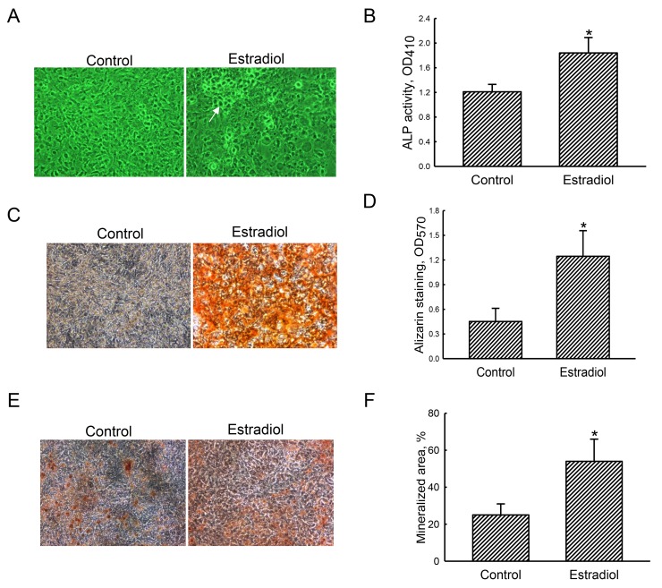
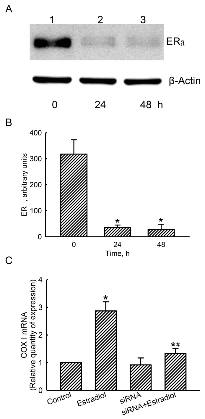
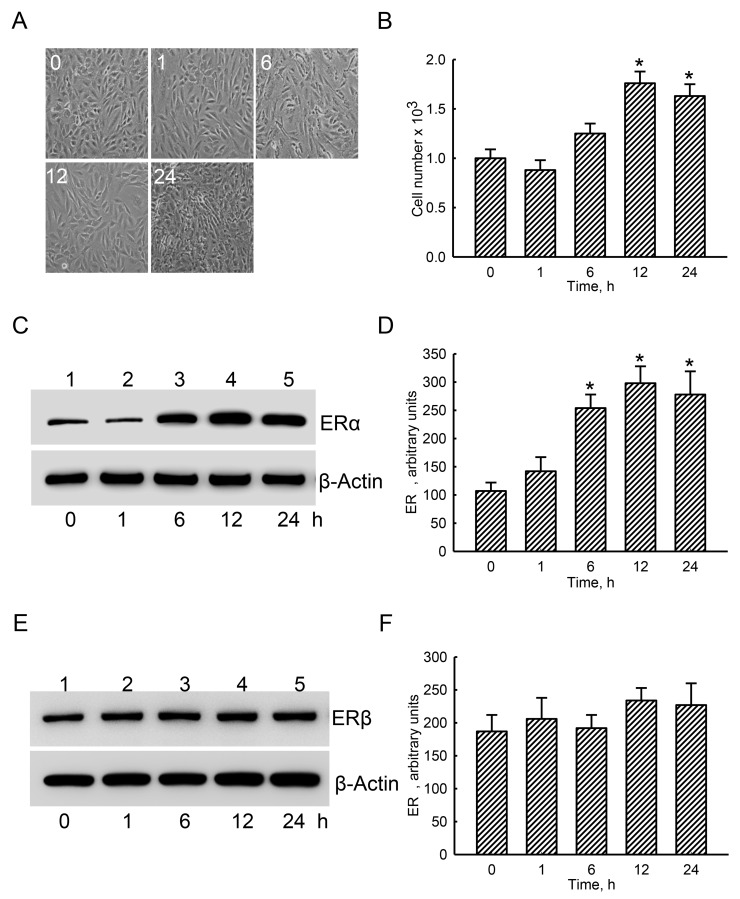
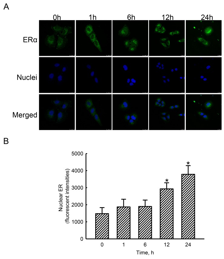
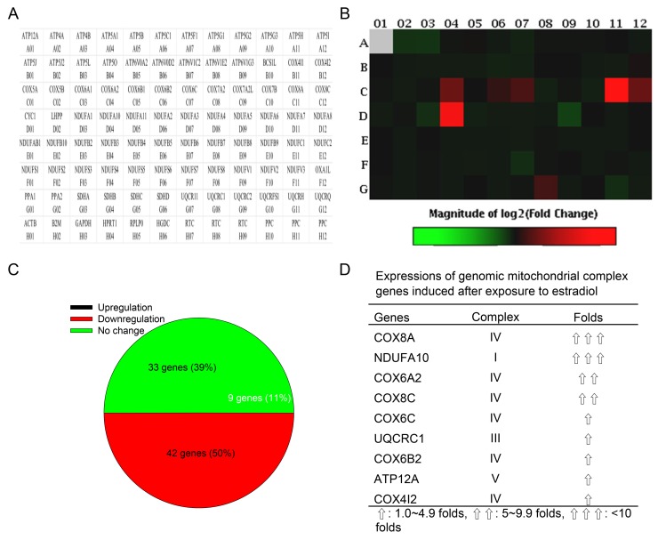
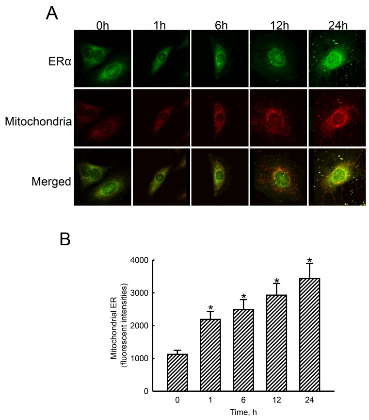
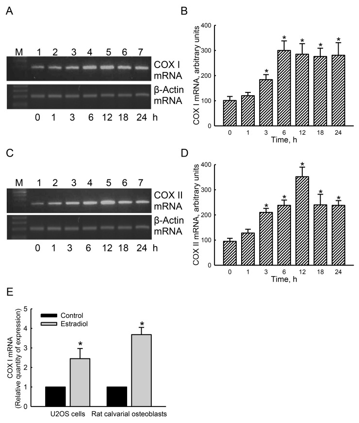
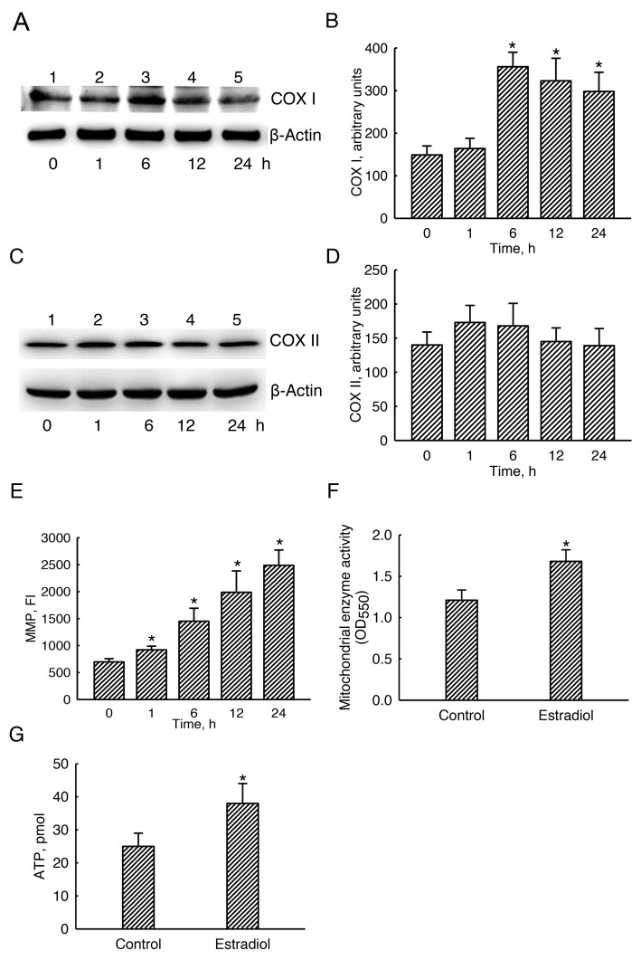
Estrogen deficiency usually leads to bone loss and osteoporosis in postmenopausal women. Osteoblasts play crucial roles in bone formation. However, osteoblast functions are influenced by mitochondrial bioenergetic conditions. In this study, we investigated the roles of the estrogen and estrogen receptor alpha (ERα) axis in mitochondrial energy metabolism and subsequent osteoblast mineralization. Exposure of rat calvarial osteoblasts to estradiol caused substantial improvements in alkaline phosphatase activities and cell calcification. In parallel, treatment of human osteoblast-like U2OS cells, derived from a female osteosarcoma patient, with estradiol specifically augmented ERα levels. Sequentially, estradiol stimulated translocation of ERα to nuclei in human osteoblasts and induced expressions of genomic respiratory chain complex , , cytochrome c oxidase (), , , , , , and genes. Concurrently, estradiol stimulated translocation of ERα to mitochondria from the cytoplasm. A bioinformatic search found the existence of four estrogen response elements in the 5'-promoter region of the mitochondrial gene. Interestingly, estradiol induced COX I mRNA and protein expressions in human osteoblasts or rat calvarial osteoblasts. Knocking-down ERα translation concurrently downregulated estradiol-induced COX I mRNA expression. Consequently, exposure to estradiol led to successive increases in the mitochondrial membrane potential, the mitochondrial enzyme activity, and cellular adenosine triphosphate levels. Taken together, this study showed the roles of the estradiol/ERα signaling axis in improving osteoblast maturation through upregulating the mitochondrial bioenergetic system due to induction of definite chromosomal and mitochondrial complex gene expressions. Our results provide novel insights elucidating the roles of the estrogen/ERα alliance in regulating bone formation.
雌激素缺乏通常会导致绝经后女性骨质流失和骨质疏松。成骨细胞在骨形成过程中发挥着关键作用。然而,成骨细胞的功能受线粒体生物能量状态的影响。在本研究中,我们探究了雌激素和雌激素受体α(ERα)轴在线粒体能量代谢及随后的成骨细胞矿化过程中的作用。将大鼠颅骨成骨细胞暴露于雌二醇可显著提高碱性磷酸酶活性和细胞钙化程度。与此同时,用雌二醇处理源自一名女性骨肉瘤患者的人成骨样U2OS细胞,可特异性提高ERα水平。随后,雌二醇刺激人成骨细胞中ERα向细胞核的转位,并诱导基因组呼吸链复合体、、细胞色素c氧化酶()、、、、和基因的表达。同时,雌二醇刺激ERα从细胞质向线粒体转位。一项生物信息学搜索发现线粒体基因的5'-启动子区域存在四个雌激素反应元件。有趣的是,雌二醇可诱导人成骨细胞或大鼠颅骨成骨细胞中COX I mRNA和蛋白的表达。敲低ERα的翻译同时会下调雌二醇诱导的COX I mRNA表达。因此,暴露于雌二醇会导致线粒体膜电位、线粒体酶活性和细胞三磷酸腺苷水平相继升高。综上所述,本研究表明雌二醇/ERα信号轴通过上调线粒体生物能量系统,诱导特定染色体和线粒体复合体基因表达,从而促进成骨细胞成熟。我们的结果为阐明雌激素/ERα联合在调节骨形成中的作用提供了新的见解。