Hsu Kuo-Sheng, Kao Hung-Ying
1Department of Biochemistry, Case Western Reserve University, 10900 Euclid Avenue, Cleveland, OH 44106 USA.
Present Address: Tumor Angiogenesis Section, Mouse Cancer Genetics Program (MCGP), National Cancer Institute (NCI), NIH, Frederick, MD 21702 USA.
Cell Biosci. 2018 Jan 25;8:5. doi: 10.1186/s13578-018-0204-8. eCollection 2018.
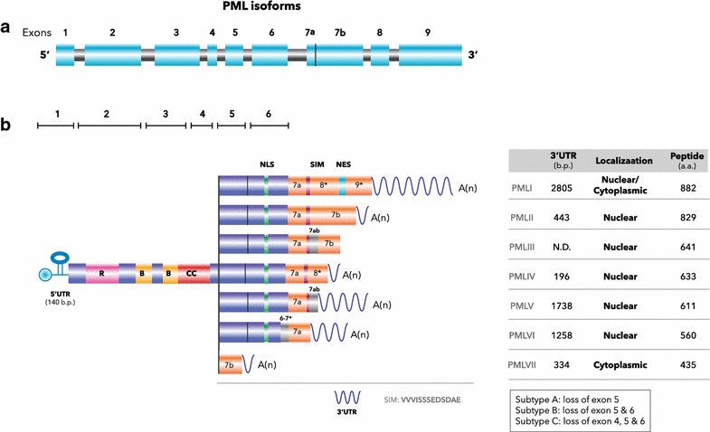
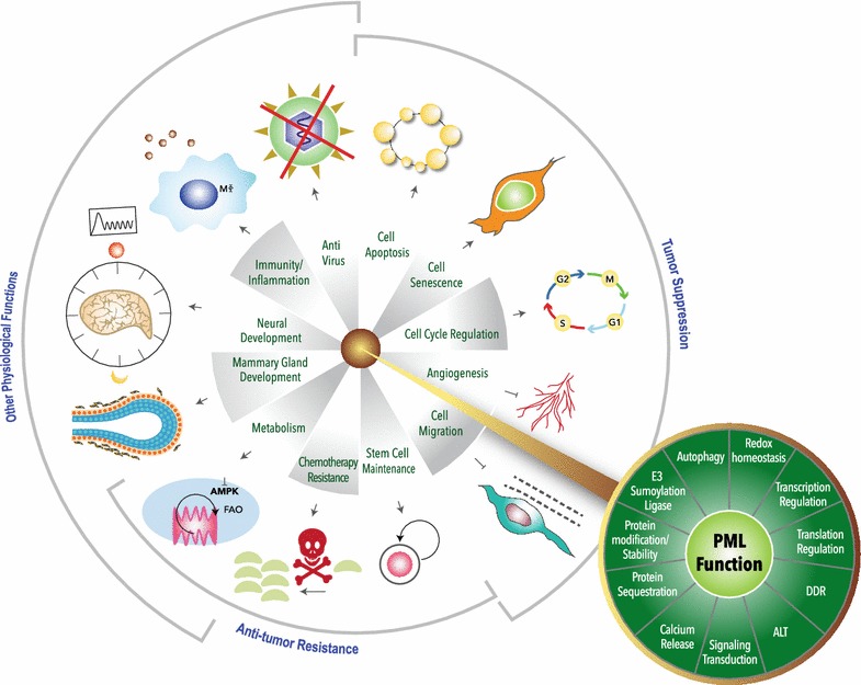
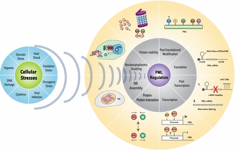
Promyelocytic leukemia protein (PML) was originally identified as a fusion partner of retinoic acid receptor alpha in acute promyelocytic leukemia patients with the (15;17) chromosomal translocation, giving rise to PML-RARα and RARα-PML fusion proteins. A body of evidence indicated that PML possesses tumor suppressing activity by regulating apoptosis, cell cycle, senescence and DNA damage responses. PML is enriched in discrete nuclear substructures in mammalian cells with 0.2-1 μm diameter in size, referred to as alternately Kremer bodies, nuclear domain 10, PML oncogenic domains or PML nuclear bodies (NBs). Dysregulation of PML NB formation results in altered transcriptional regulation, protein modification, apoptosis and cellular senescence. In addition to PML NBs, PML is also present in nucleoplasm and cytoplasmic compartments, including the endoplasmic reticulum and mitochondria-associated membranes. The role of PML in tumor suppression has been extensively studied but increasing evidence indicates that PML also plays versatile roles in stem cell renewal, metabolism, inflammatory responses, neural function, mammary development and angiogenesis. In this review, we will briefly describe the known PML regulation and function and include new findings.
早幼粒细胞白血病蛋白(PML)最初是在患有(15;17)染色体易位的急性早幼粒细胞白血病患者中被鉴定为维甲酸受体α的融合伴侣,从而产生PML-RARα和RARα-PML融合蛋白。大量证据表明,PML通过调节细胞凋亡、细胞周期、衰老和DNA损伤反应而具有肿瘤抑制活性。PML在哺乳动物细胞中富集于直径为0.2-1μm的离散核亚结构中,这些亚结构被交替称为克雷默小体、核域10、PML致癌结构域或PML核体(NBs)。PML核体形成的失调会导致转录调控、蛋白质修饰、细胞凋亡和细胞衰老的改变。除了PML核体,PML还存在于核质和细胞质区室中,包括内质网和线粒体相关膜。PML在肿瘤抑制中的作用已得到广泛研究,但越来越多的证据表明,PML在干细胞更新、代谢、炎症反应、神经功能、乳腺发育和血管生成中也发挥着多种作用。在这篇综述中,我们将简要描述已知的PML调控和功能,并纳入新发现。