Kearney Hugh, Cryan Jane, Beausang Alan, Looby Seamus, Brett Francesca M
Clin Neuropathol. 2018 May/Jun;37(3):97-104. doi: 10.5414/NP301084.
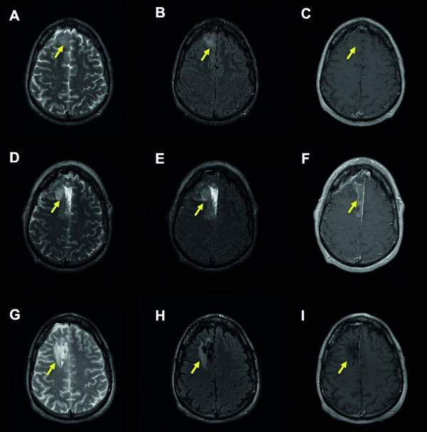
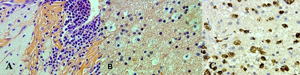
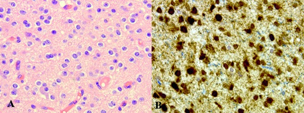
The aim of this study is to identify, in our center, all cases of foreign-body reactions to hemostatic agents or other prostheses resulting in a radiological suspicion of tumor recurrence. We interrogated our internal database to identify all such cases and systematically evaluated the MRI brain scans of patients: (i) at the time of initial tumor diagnosis, (ii) postoperatively, (iii) and at the time of suspected tumor recurrence. In addition, we reviewed each patient's operative notes and reviewed the histology of all cases following a second surgical intervention. In total, we identified 8 patients, 7 of whom had a WHO grade II glioma at initial surgery. We did not identify any distinguishing radiological abnormalities from the initial diagnostic brain scan to the suspected recurrence, and histologically all cases were characterized by extensive gliosis; with both macrophages and reactive astrocytes present throughout. The cause of gliosis was identified as being relating to hemostatic agents in 4 cases; in the other 4 cases, the foreign-body reaction was presumed to be caused be materials used in a craniotomy or cranioplasty. This study highlights the difficulty in radiologically diagnosing a foreign-body reaction and also identifies that such a gliotic reaction may occur as a consequence of exogenous materials used in a craniotomy or cranioplasty. .
本研究的目的是在我们中心识别所有因对止血剂或其他假体产生异物反应而导致影像学怀疑肿瘤复发的病例。我们查询了内部数据库以识别所有此类病例,并系统地评估了患者的脑部MRI扫描:(i)在最初肿瘤诊断时,(ii)术后,以及(iii)在怀疑肿瘤复发时。此外,我们查看了每位患者的手术记录,并在二次手术干预后复查了所有病例的组织学情况。我们总共识别出8例患者,其中7例在初次手术时患有世界卫生组织二级胶质瘤。从最初的诊断性脑部扫描到疑似复发,我们未发现任何明显的影像学异常,并且从组织学上看,所有病例均以广泛的胶质增生为特征;整个过程中均存在巨噬细胞和反应性星形胶质细胞。在4例病例中,胶质增生的原因被确定与止血剂有关;在其他4例病例中,异物反应被推测是由开颅手术或颅骨成形术中使用的材料引起的。这项研究凸显了在影像学上诊断异物反应的困难,同时也表明这种胶质反应可能是开颅手术或颅骨成形术中使用的外源材料所致。