Department of Orthodontics, Affiliated Hospital of Stomatology, Medical College, Zhejiang University, Hangzhou, Zhejiang Province, China.
Department of Liver Surgery, Ren Ji Hospital Affiliated to Shanghai Jiao Tong University School of Medicine, Shanghai, China.
Cell Death Dis. 2018 Feb 12;9(2):207. doi: 10.1038/s41419-018-0279-5.
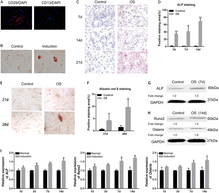
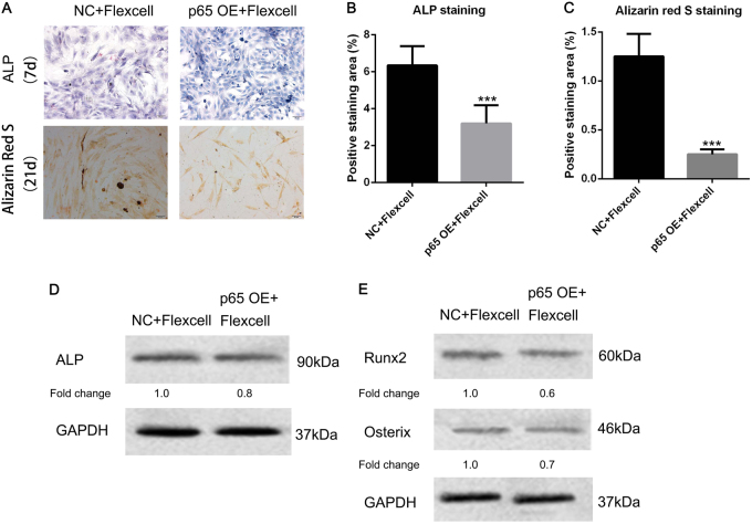
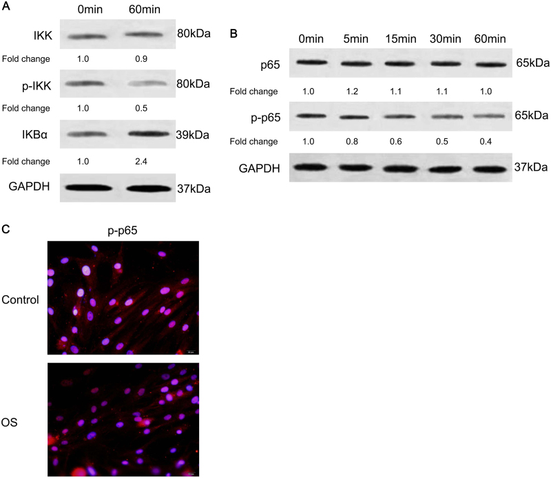
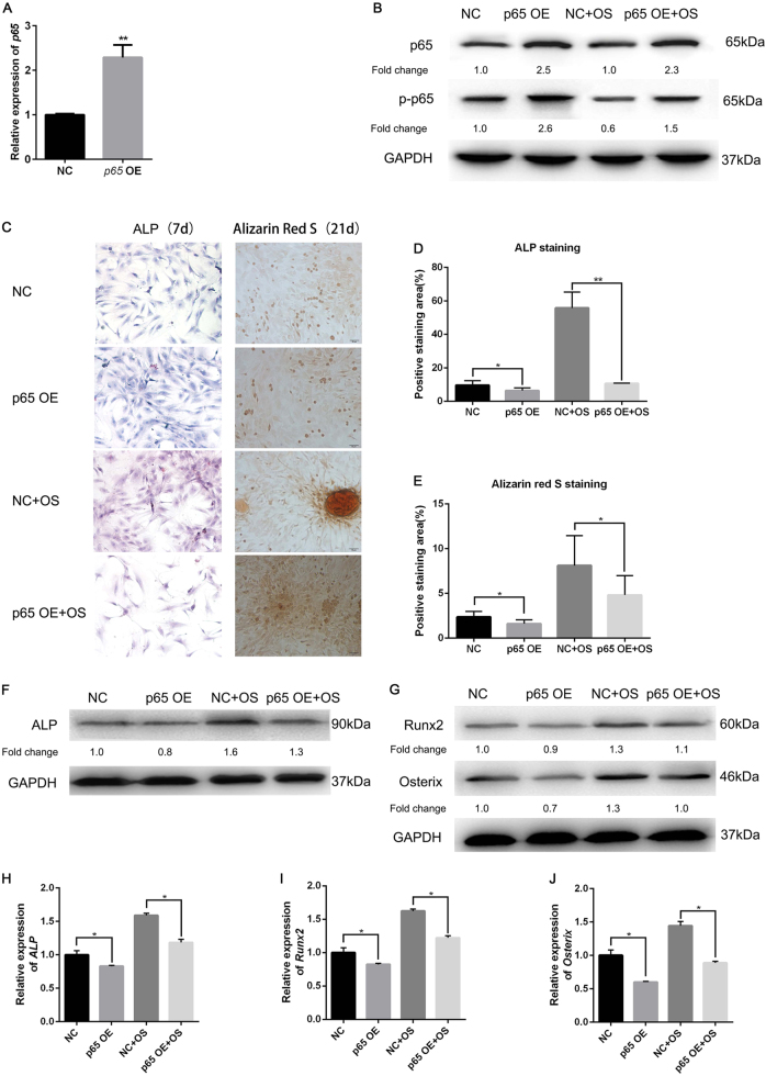
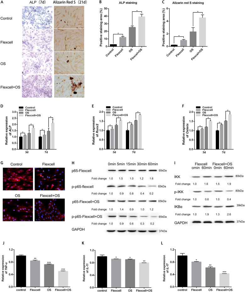
Severe malocclusion can contribute to several serious dental and physical conditions, such as digestive difficulties, periodontal disease, and severe tooth decay. Orthodontic treatment is mainly used to treat malocclusion. Forces in orthodontic tooth results in bone resorption on the pressure side and bone deposition on the tension side. Osteoblasts have been considered as the key component in bone regeneration on the tension side. However, the underlying mechanisms remain unclear. In this study, we focus on how mechanical stretch regulates the osteogenesis during orthodontic treatment. Human jaw bone marrow mesenchymal stem cells (hJBMMSCs) were isolated from healthy adult donors and cultured in regular medium (control) or osteogenic medium (OS). Under OS culture, hJBMMSCs presented osteogenic differentiation potentials, as evidenced by increased mineralization, enhanced calcium deposition, and upregulated expression of osteogenesis markers (ALP, osterix, and Runx). What's more, the OS-induced osteogenesis of hJBMMSCs is associated with the dephosphorylation of IKK, activation of IKBα, and phosphorylation/nucleic accumulation of P65, which all indicated the inhibition of NF-κB activity. Overexpressing P65 in hJBMMSCs, which could constantly activate NF-κB, prevented the osteogenic differentiation in the OS. After that, we applied the Flexcell tension system, which could cause mechanical stretch on cultured hJBMMSCs to mimic the tension forces during tooth movement. Mechanical stretch resulted in 3.5-fold increase of ALP activity and 2.4-fold increase of calcium deposition after 7 days and 21 days treatment, respectively. The expression levels of ALP, Run×2, and Osterix were also significantly upregulated. In the meantime, applying mechanical stretch on OS-cultured hJBMMSCs also dramatically promoted the OS-induced osteogenesis. Both OS and mechanical stretch downregulated NF-κB activity. By overexpressing P65 in hJBMMSCs, neither OS nor mechanical stretch could induce their osteogenesis. These results indicated that, like OS induction, mechanical stretch-facilitated osteogenesis of hJBMMSCs by inhibiting NF-κB in the noninflammatory environments.
严重的错颌畸形会导致许多严重的牙齿和身体状况,如消化困难、牙周病和严重的蛀牙。正畸治疗主要用于治疗错颌畸形。正畸牙齿的力会导致压力侧的骨吸收和张力侧的骨沉积。成骨细胞被认为是张力侧骨再生的关键组成部分。然而,其潜在机制尚不清楚。在这项研究中,我们专注于机械拉伸如何调节正畸治疗过程中的成骨作用。从健康成年供体中分离出人颌骨髓间充质干细胞(hJBMMSCs),并在常规培养基(对照)或成骨培养基(OS)中培养。在 OS 培养下,hJBMMSCs 表现出成骨分化潜能,表现为矿化增加、钙沉积增强以及成骨标志物(ALP、osterix 和 Runx)的表达上调。更重要的是,OS 诱导的 hJBMMSCs 成骨作用与 IKK 的去磷酸化、IKBα 的激活以及 P65 的磷酸化/核积累有关,这都表明 NF-κB 活性受到抑制。在 hJBMMSCs 中转染 P65 可持续激活 NF-κB,从而阻止 OS 中的成骨分化。之后,我们应用 Flexcell 张力系统对培养的 hJBMMSCs 施加机械拉伸,以模拟牙齿移动过程中的张力。机械拉伸分别在 7 天和 21 天处理后导致 ALP 活性增加 3.5 倍,钙沉积增加 2.4 倍。ALP、Run×2 和 Osterix 的表达水平也显著上调。同时,在 OS 培养的 hJBMMSCs 上施加机械拉伸也显著促进了 OS 诱导的成骨作用。OS 和机械拉伸均降低了 NF-κB 的活性。在 hJBMMSCs 中转染 P65 后,OS 和机械拉伸均不能诱导其成骨作用。这些结果表明,与 OS 诱导一样,机械拉伸在非炎症环境中通过抑制 NF-κB 促进 hJBMMSCs 的成骨作用。