Bishop Courtney A, Ricotti Valeria, Sinclair Christopher D J, Evans Matthew R B, Butler Jordan W, Morrow Jasper M, Hanna Michael G, Matthews Paul M, Yousry Tarek A, Muntoni Francesco, Thornton John S, Newbould Rexford D, Janiczek Robert L
Imanova Limited, Hammersmith Hospital, London, United Kingdom.
Dubowitz Neuromuscular Centre, UCL Great Ormond Street Institute of Child Health, London, United Kingdom.
Front Neurol. 2018 Jan 26;9:9. doi: 10.3389/fneur.2018.00009. eCollection 2018.
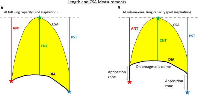
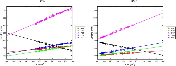
Subjects with Duchenne Muscular Dystrophy (DMD) suffer from progressive muscle damage leading to diaphragmatic weakness that ultimately requires ventilation. Emerging treatments have generated interest in better characterizing the natural history of respiratory impairment in DMD and responses to therapy. Dynamic (cine) Magnetic Resonance Imaging (MRI) may provide a more sensitive measure of diaphragm function in DMD than the commonly used spirometry. This study presents an analysis pipeline for measuring parameters of diaphragmatic motion from dynamic MRI and its application to investigate MRI measures of respiratory function in both healthy controls and non-ambulant DMD boys. We scanned 13 non-ambulant DMD boys and 10 age-matched healthy male volunteers at baseline, with a subset ( = 10, 10, 8) of the DMD subjects also assessed 3, 6, and 12 months later. Spirometry-derived metrics including forced vital capacity were recorded. The MRI-derived measures included the lung cross-sectional area (CSA), the anterior, central, and posterior lung lengths in the sagittal imaging plane, and the diaphragm length over the time-course of the dynamic MRI. Regression analyses demonstrated strong linear correlations between lung CSA and the length measures over the respiratory cycle, with a reduction of these correlations in DMD, and diaphragmatic motions that contribute less efficiently to changing lung capacity in DMD. MRI measures of pulmonary function were reduced in DMD, controlling for height differences between the groups: at maximal inhalation, the maximum CSA and the total distance of motion of the diaphragm were 45% and 37% smaller. MRI measures of pulmonary function were correlated with spirometry data and showed relationships with disease progression surrogates of age and months non-ambulatory, suggesting that they provide clinically meaningful information. Changes in the MRI measures over 12 months were consistent with weakening of diaphragmatic and inter-costal muscles and progressive diaphragm dysfunction. In contrast, longitudinal changes were not seen in conventional spirometry measures during the same period. Dynamic MRI measures of thoracic muscle and pulmonary function are, therefore, believed to detect meaningful differences between healthy controls and DMD and may be sensitive to changes in function over relatively short periods of follow-up in non-ambulant boys with DMD.
杜氏肌营养不良症(DMD)患者会遭受进行性肌肉损伤,导致膈肌无力,最终需要通气。新兴疗法引发了人们对更好地描述DMD呼吸功能损害的自然史以及对治疗反应的兴趣。动态(电影)磁共振成像(MRI)可能比常用的肺活量测定法更能敏感地测量DMD患者的膈肌功能。本研究提出了一种用于从动态MRI测量膈肌运动参数的分析流程,并将其应用于研究健康对照和非行走型DMD男孩的呼吸功能MRI测量。我们在基线时扫描了13名非行走型DMD男孩和10名年龄匹配的健康男性志愿者,其中一部分DMD受试者(分别为10名、10名、8名)在3个月、6个月和12个月后也进行了评估。记录了包括用力肺活量在内的肺活量测定指标。MRI测量指标包括肺横截面积(CSA)、矢状面成像平面上肺的前、中、后长度,以及动态MRI过程中膈肌的长度。回归分析表明,肺CSA与呼吸周期中的长度测量值之间存在强线性相关性,在DMD患者中这些相关性降低,并且DMD患者中膈肌运动对改变肺容量的效率较低。在控制了两组之间的身高差异后,DMD患者的肺功能MRI测量值降低:在最大吸气时,最大CSA和膈肌运动的总距离分别小45%和37%。肺功能的MRI测量值与肺活量测定数据相关,并与年龄和非行走月数等疾病进展替代指标相关,表明它们提供了具有临床意义的信息。12个月内MRI测量值的变化与膈肌和肋间肌的减弱以及膈肌功能的进行性障碍一致。相比之下,同期传统肺活量测定指标未见纵向变化。因此,胸肌和肺功能的动态MRI测量被认为能够检测出健康对照和DMD之间的有意义差异,并且可能对非行走型DMD男孩在相对较短的随访期内的功能变化敏感。