Zhao Bingxia, Chen Yihan, Liu Jinfeng, Zhang Li, Wang Jing, Yang Yali, Lv Qing, Xie Mingxing
Department of Ultrasound, Union Hospital, Tongji Medical College, Huazhong University of Science and Technology, Wuhan 430022, China.
Hubei Key Laboratory of Molecular Imaging, Union Hospital, Tongji Medical College, Huazhong University of Science and Technology, Wuhan 430022, China.
Oncotarget. 2017 Dec 21;9(4):4897-4914. doi: 10.18632/oncotarget.23527. eCollection 2018 Jan 12.
To investigate the effects of the microbubble (MB) dose, mechanism index (MI) and sonication duration on blood-brain barrier (BBB) disruption induced by diagnostic ultrasound combined with MBs as well as to investigate the potential molecular mechanism.
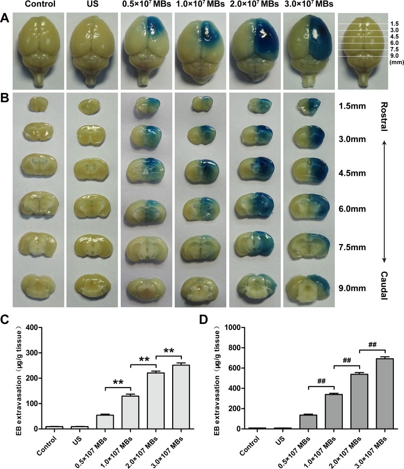
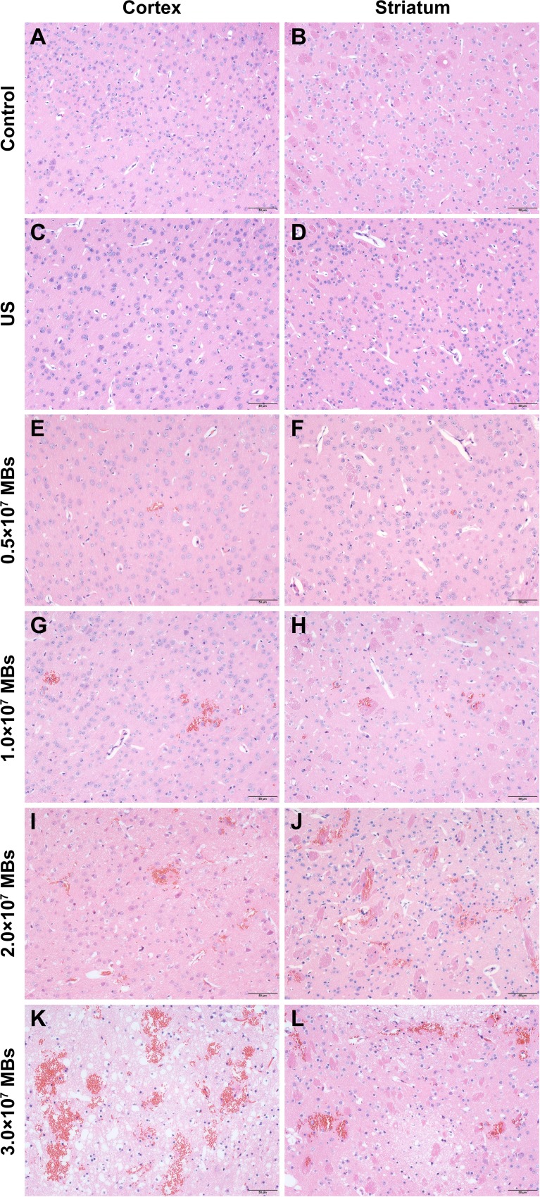
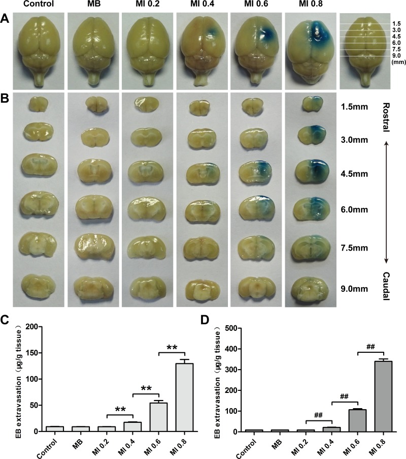
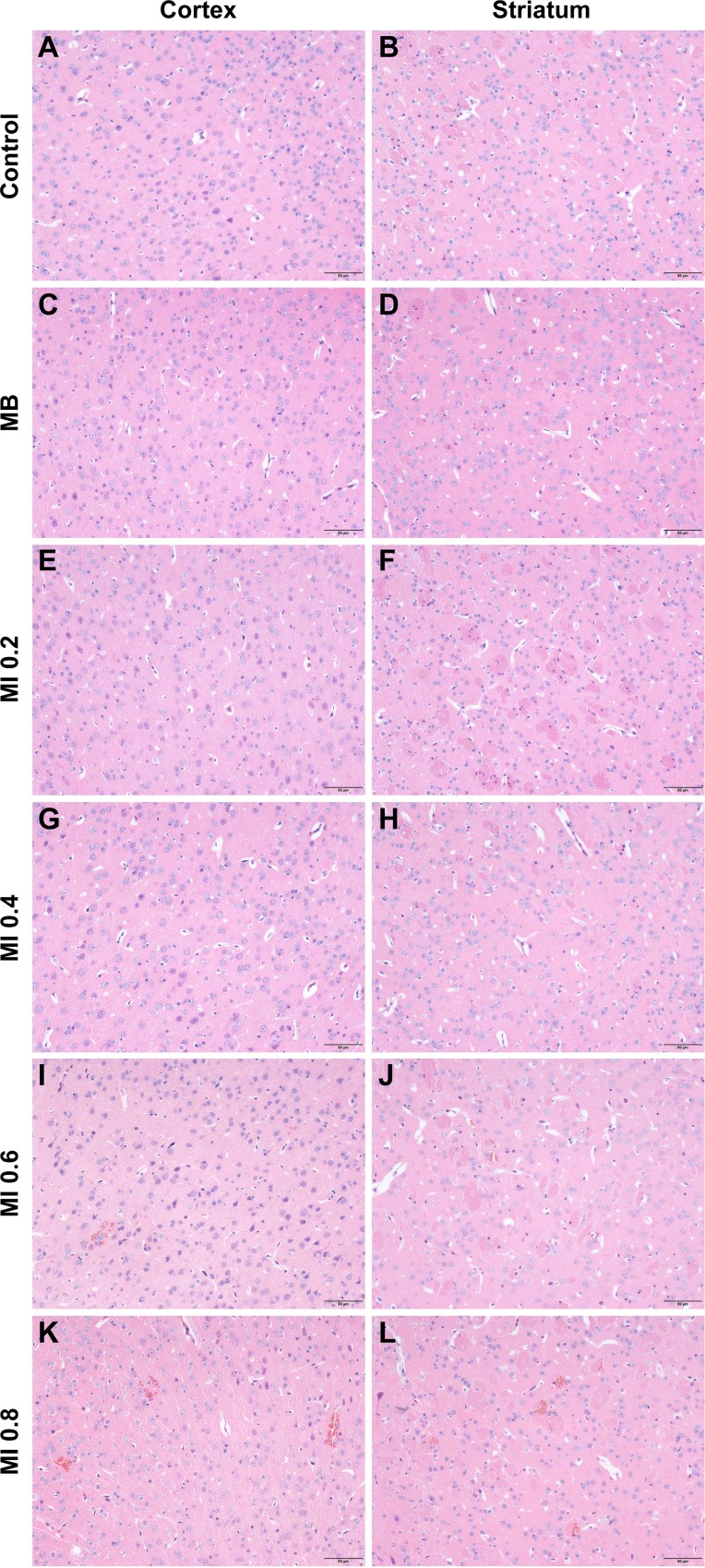
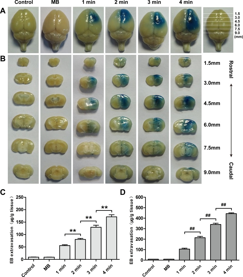
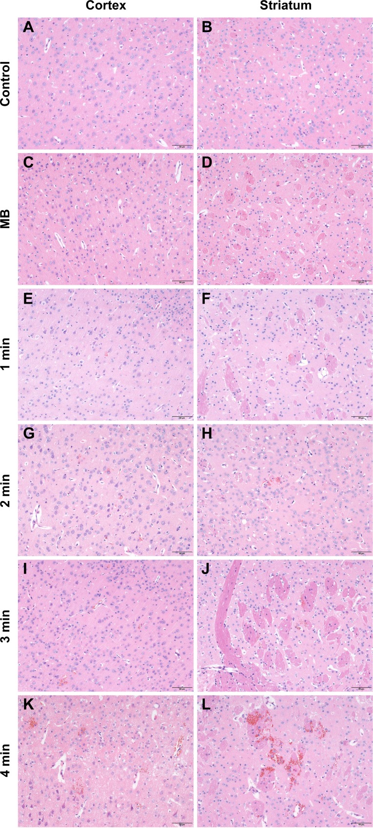
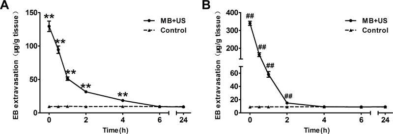
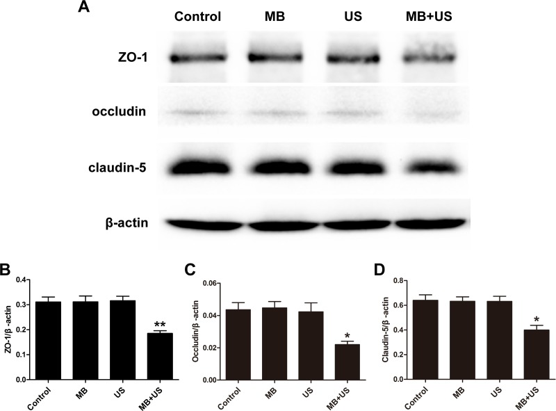
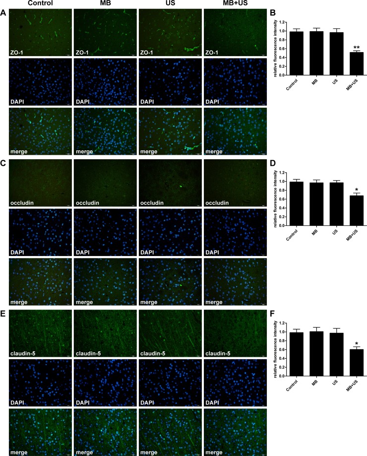
The extent of BBB disruption increased with MB dose, MI and sonication duration. A relatively larger extent of BBB disruption associated with minimal tissue damage was achieved by an appropriate MB dose and ultrasound exposure parameters with diagnostic ultrasound. Decreased expression of ZO-1, occludin and claudin-5 were correlated with disruption of the BBB, as confirmed by paracellular passage of the tracer lanthanum nitrate into the brain parenchyma after BBB disruption.
These findings indicated that this technique is a promising tool for promoting brain delivery of diagnostic and therapeutic agents in the diagnosis and treatment of brain diseases.
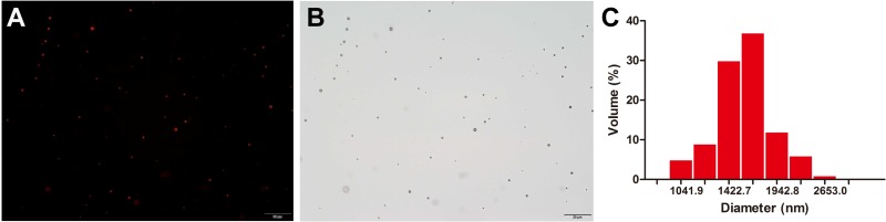
The extent of BBB disruption was qualitatively assessed by Evans blue (EB) staining and quantitatively analyzed by an EB extravasation measurement. A histological examination was performed to evaluate tissue damage. Expression of tight junction (TJ) related proteins ZO-1, occludin and claudin-5 was determined by western blotting analysis and immunohistofluorescence. Transmission electron microscopy was performed to observe ultrastructure changes of TJs after BBB disruption.
研究微泡(MB)剂量、机械指数(MI)和超声照射时间对诊断超声联合微泡诱导的血脑屏障(BBB)破坏的影响,并探讨其潜在的分子机制。
血脑屏障破坏程度随微泡剂量、机械指数和超声照射时间的增加而增加。通过适当的微泡剂量和诊断超声的超声暴露参数,可在组织损伤最小的情况下实现相对较大程度的血脑屏障破坏。血脑屏障破坏后,示踪剂硝酸镧经细胞旁通道进入脑实质,证实紧密连接蛋白1(ZO-1)、闭合蛋白和Claudin-5表达降低与血脑屏障破坏相关。
这些发现表明,该技术是一种有前景的工具,可用于在脑部疾病的诊断和治疗中促进诊断和治疗药物的脑内递送。
通过伊文思蓝(EB)染色定性评估血脑屏障破坏程度,并通过EB外渗测量进行定量分析。进行组织学检查以评估组织损伤。通过蛋白质免疫印迹分析和免疫荧光法测定紧密连接(TJ)相关蛋白ZO-1、闭合蛋白和Claudin-5的表达。进行透射电子显微镜检查以观察血脑屏障破坏后紧密连接的超微结构变化。