Li Ching-Wen, Davis Brett, Shea Jill, Sant Himanshu, Gale Bruce Kent, Agarwal Jayant
Department of Mechanical Engineering, National Chung Hsing University, Taichung, Taiwan, China.
Department of Surgery, School of Medicine, University of Utah, Salt Lake City, UT, USA.
Neural Regen Res. 2018 Jan;13(1):105-111. doi: 10.4103/1673-5374.224377.
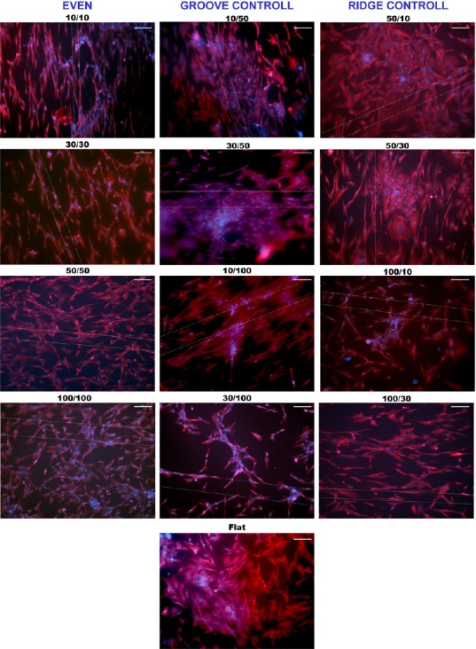
Nerve conduits have been a viable alternative to the 'gold standard' autograft for treating small peripheral nerve gap injuries. However, they often produce inadequate functional recovery outcomes and are ineffective in large gap injuries. Ridge/groove surface micropatterning has been shown to promote neural cell orientation and guide growth. However, optimization of the ratio of ridge/groove parameters to promote orientation and extension for dorsal root ganglion (DRG) cells on poly(lactic-co-glycolic acid) (PLGA) films has not been previously conducted. Photolithography and micro-molding were used to define various combinations of ridge/groove dimensions on PLGA films. The DRG cells obtained from chicken embryos were cultured on micropatterned PLGA films for cell orientation and migration evaluation. Biodegradation of the films occurred during the test period, however, this did not cause deformation or distortion of the micropatterns. Results from the DRG cell orientation test suggest that when the ridge/groove ratio equals 1 (ridge/groove width parameters are equal, i.e., 10 μm/10 μm (even)), the degree of alignment depends on the size of the ridges and grooves, when the ratio is smaller than 1 (groove controlled) the alignment increases as the ridge size decreases, and when the ratio is larger than 1 (ridge controlled), the alignment is reduced as the width of the grooves decreases. The migration rate and neurite extension of DRG neurons were greatest on 10 μm/10 μm and 30 μm/30 μm micropatterned PLGA films. Based on the data, the 10 μm/10 μm and 30 μm/30 μm micropatterned PLGA films are the optimized ridge/groove surface patterns for the construction of nerve repair devices.
神经导管已成为治疗小面积周围神经间隙损伤的“金标准”自体移植物的可行替代方案。然而,它们往往产生的功能恢复效果不佳,并且在大面积间隙损伤中无效。脊/槽表面微图案化已被证明可促进神经细胞定向并引导生长。然而,以前尚未对聚乳酸-乙醇酸共聚物(PLGA)薄膜上促进背根神经节(DRG)细胞定向和延伸的脊/槽参数比例进行优化。使用光刻和微成型技术在PLGA薄膜上定义各种脊/槽尺寸组合。将从鸡胚胎获得的DRG细胞培养在微图案化的PLGA薄膜上,以评估细胞定向和迁移。在测试期间薄膜发生了生物降解,然而,这并未导致微图案变形或扭曲。DRG细胞定向测试结果表明,当脊/槽比例等于1(脊/槽宽度参数相等,即10μm/10μm(偶数))时,排列程度取决于脊和槽的大小;当比例小于1(槽控制)时,随着脊尺寸减小排列增加;当比例大于1(脊控制)时,随着槽宽度减小排列减少。DRG神经元在10μm/10μm和30μm/30μm微图案化PLGA薄膜上的迁移率和神经突延伸最大。基于这些数据,10μm/10μm和30μm/30μm微图案化PLGA薄膜是用于构建神经修复装置的优化脊/槽表面图案。