Department of Neuroimmunology, Center for Brain Research, Medical University of Vienna, Vienna.
Clinical Department of Neurology, Medical University of Innsbruck, Innsbruck, Austria.
Curr Opin Neurol. 2018 Jun;31(3):325-333. doi: 10.1097/WCO.0000000000000551.
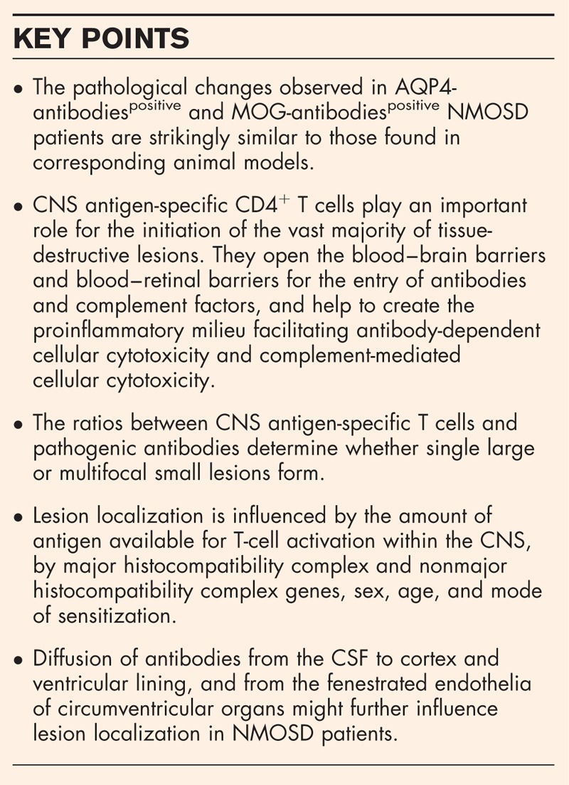
Neuromyelitis optica spectrum disorders (NMOSD) are severe inflammatory diseases of the central nervous system (CNS), with the presence of aquaporin 4 (AQP4)-specific serum antibodies in the vast majority of patients, and with the presence of myelin oligodendrocyte glycoprotein (MOG)-specific antibodies in approximately 40% of all AQP4-antibody negative NMOSD patients. Despite differences in antigen recognition, the preferred sites of lesions are similar in both groups of patients: They localize to the spinal cord and to the anterior visual pathway including retina, optic nerves, chiasm, and optic tracts, and - to lesser extent - also to certain predilection sites in the brain.
The involvement of T cells in the formation of NMOSD lesions has been challenged for quite some time. However, several recent findings demonstrate the key role of T cells for lesion formation and localization. Studies on the evolution of lesions in the spinal cord of NMOSD patients revealed a striking similarity of early NMOSD lesions with those observed in corresponding T-cell-induced animal models, both in lesion formation and in lesion localization. Studies on retinal abnormalities in NMOSD patients and corresponding animals revealed the importance of T cells for the very early stages of retinal lesions which eventually culminate in damage to Müller cells and to the retinal nerve fiber layer. Finally, a study on cerebrospinal fluid (CSF) barrier pathology demonstrated that NMOSD immunopathology extends beyond perivascular astrocytic foot processes to include the pia, the ependyma, and the choroid plexus, and that diffusion of antibodies from the CSF could further influence lesion formation in NMOSD patients.
The pathological changes observed in AQP4-antibody positive and MOG-antibody positive NMOSD patients are strikingly similar to those found in corresponding animal models, and many mechanisms which determine lesion localization in experimental animals seem to closely reflect the human situation.
视神经脊髓炎谱系疾病(NMOSD)是一种严重的中枢神经系统(CNS)炎症性疾病,绝大多数患者存在水通道蛋白 4(AQP4)特异性血清抗体,约 40%的所有 AQP4 抗体阴性 NMOSD 患者存在髓鞘少突胶质细胞糖蛋白(MOG)特异性抗体。尽管抗原识别存在差异,但两组患者的病变部位相似:病变定位于脊髓和前视路,包括视网膜、视神经、视交叉和视束,且在一定程度上也定位于大脑的某些易患部位。
T 细胞在 NMOSD 病变形成中的作用已有一段时间受到质疑。然而,一些最近的发现表明 T 细胞在病变形成和定位中起着关键作用。对 NMOSD 患者脊髓病变演变的研究表明,NMOSD 早期病变与相应 T 细胞诱导的动物模型中观察到的病变非常相似,无论是在病变形成还是在病变定位方面。对 NMOSD 患者和相应动物的视网膜异常的研究揭示了 T 细胞对视网膜病变早期阶段的重要性,最终导致 Müller 细胞和视网膜神经纤维层受损。最后,一项关于脑脊液(CSF)屏障病理学的研究表明,NMOSD 免疫病理学不仅局限于血管周围星形胶质细胞足突,还包括软膜、室管膜和脉络丛,抗体从 CSF 扩散可能进一步影响 NMOSD 患者的病变形成。
AQP4 抗体阳性和 MOG 抗体阳性 NMOSD 患者的病理变化与相应动物模型中发现的变化非常相似,许多决定实验动物病变定位的机制似乎与人类情况密切相关。