Niedenberger Bryan A, Geyer Christopher B
East Carolina Diabetes and Obesity Institute East Carolina University, Greenville, NC, USA.
East Carolina Diabetes and Obesity Institute East Carolina University, Greenville, NC, USA; Brody School of Medicine at East Carolina University, Greenville, NC, USA.
Stem Cell Res. 2018 Mar;27:162-168. doi: 10.1016/j.scr.2018.01.031. Epub 2018 Feb 9.
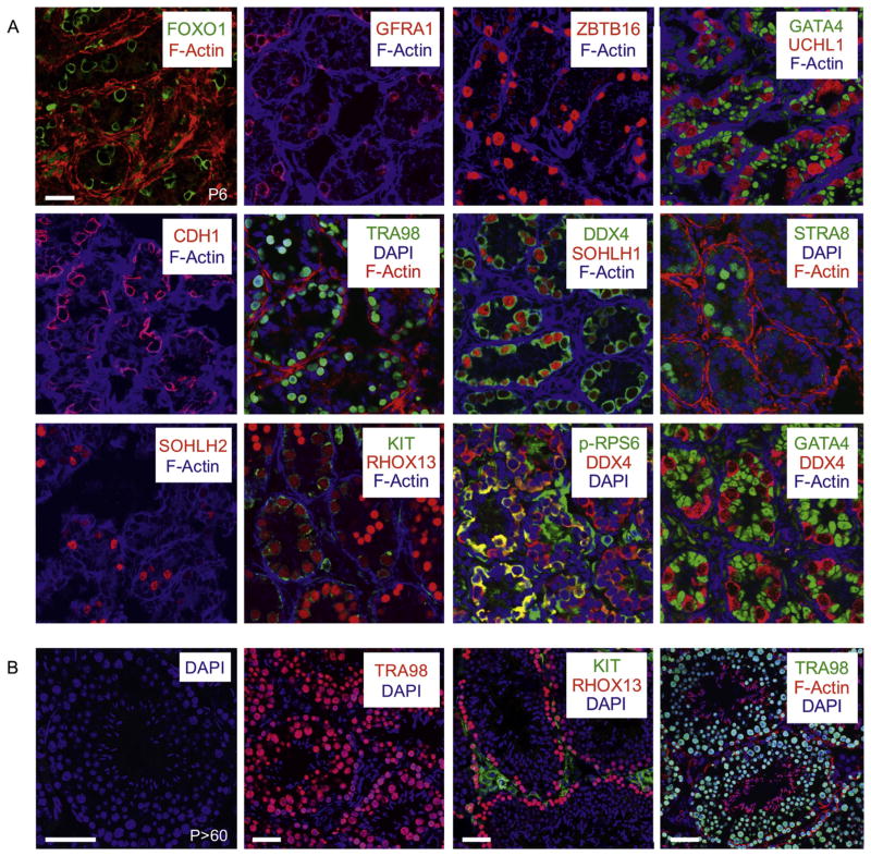
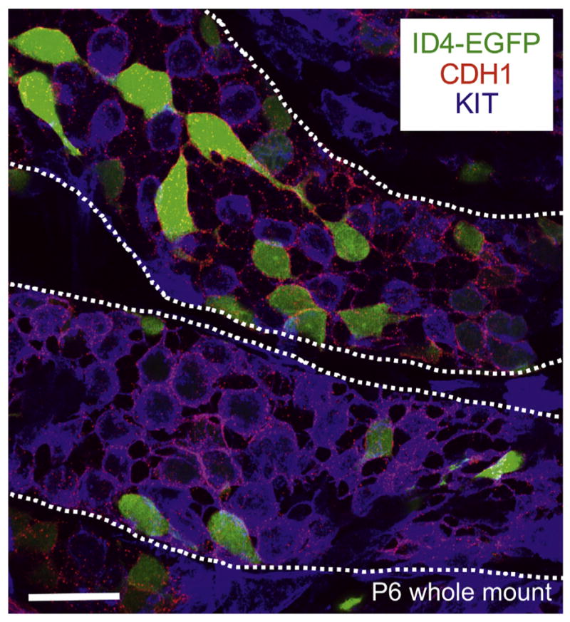
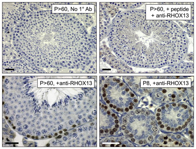
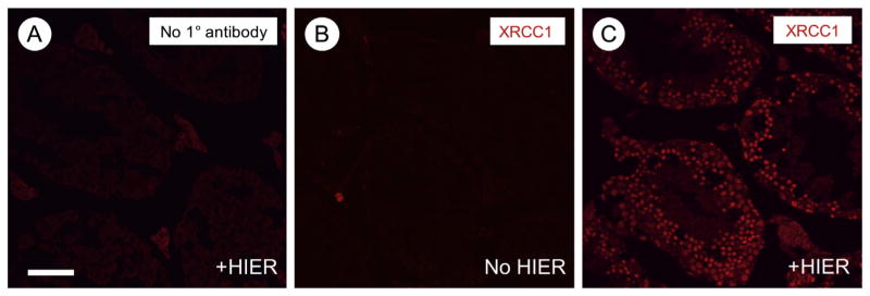
Mammalian male germ cell development takes place in the testis under the influence of a variety of somatic cells and an incompletely defined paracrine and endocrine influences. Since it is not recapitulated well in vitro, researchers studying spermatogenesis often manipulate the germline by creating transgenic or knockout mice or by administering pharmaceutical agonists/antagonists or inhibitors. The effects of these types of manipulations on germline development can often be determined following microscopic imaging, both of stained and immunostained testis sections. Here, we describe approaches for microscopic analysis of the developing male germline, provide detailed protocols for a variety of immunostaining approaches, and discuss transgenic fluorescent reporter lines for studying the early stages of spermatogenesis.
哺乳动物雄性生殖细胞的发育在多种体细胞以及尚不明确的旁分泌和内分泌影响下于睾丸中进行。由于其在体外难以很好地重现,研究精子发生的科研人员常常通过创建转基因或基因敲除小鼠,或施用药物激动剂/拮抗剂或抑制剂来操控生殖系。通过对染色和免疫染色的睾丸切片进行显微成像,往往能够确定这些类型的操控对生殖系发育的影响。在此,我们描述了发育中的雄性生殖系显微分析方法,提供了多种免疫染色方法的详细方案,并讨论了用于研究精子发生早期阶段的转基因荧光报告品系。