Department of Physics, University of Helsinki, Helsinki, Finland.
Helsinki University Central Hospital Medical Imaging Center, Helsinki, Finland.
Sci Rep. 2018 Feb 23;8(1):3519. doi: 10.1038/s41598-018-20986-x.
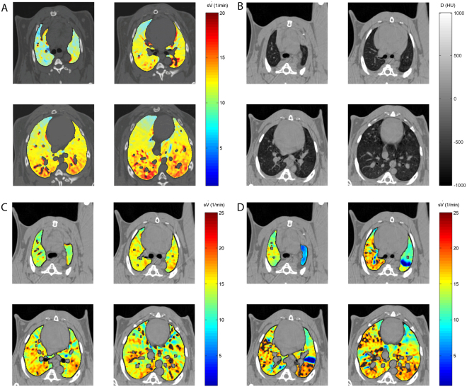
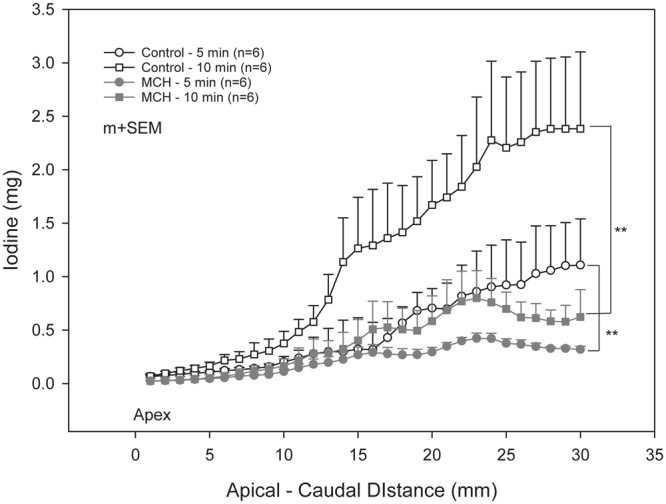
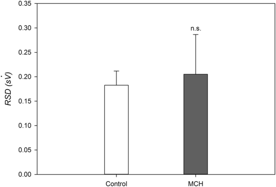
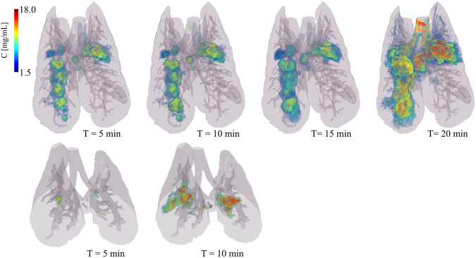
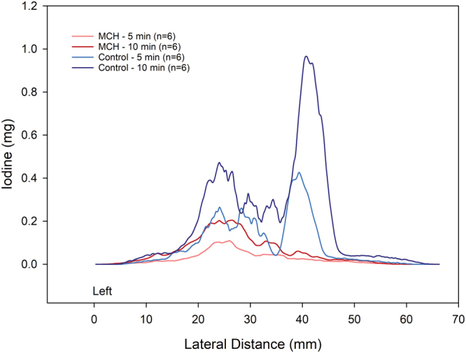
To understand the determinants of inhaled aerosol particle distribution and targeting in the lung, knowledge of regional deposition, lung morphology and regional ventilation, is crucial. No single imaging modality allows the acquisition of all such data together. Here we assessed the feasibility of dual-energy synchrotron radiation imaging to this end in anesthetized rabbits; both in normal lung (n = 6) and following methacholine (MCH)-induced bronchoconstriction (n = 6), a model of asthma. We used K-edge subtraction CT (KES) imaging to quantitatively map the regional deposition of iodine-containing aerosol particles. Morphological and regional ventilation images were obtained, followed by quantitative regional iodine deposition maps, after 5 and 10 minutes of aerosol administration. Iodine deposition was markedly inhomogeneous both in normal lung and after induced bronchoconstrition. Deposition was significantly reduced in the MCH group at both time points, with a strong dependency on inspiratory flow in both conditions (R = 0.71; p < 0.0001). We demonstrate for the first time, the feasibility of KES CT for quantitative imaging of lung deposition of aerosol particles, regional ventilation and morphology. Since these are among the main factors determining lung aerosol deposition, we expect this imaging approach to bring new contributions to the understanding of lung aerosol delivery, targeting, and ultimately biological efficacy.
为了理解吸入气溶胶颗粒在肺部的分布和靶向性的决定因素,了解区域性沉积、肺形态和区域性通气至关重要。没有单一的成像方式可以同时获取所有这些数据。在这里,我们评估了双能同步辐射成像在这方面的可行性,在麻醉兔中进行了研究;包括正常肺(n=6)和乙酰甲胆碱(MCH)诱导的支气管收缩(n=6),即哮喘模型。我们使用 K 边差分 CT(KES)成像来定量映射含碘气溶胶颗粒的区域性沉积。在给予气溶胶后 5 分钟和 10 分钟,进行形态学和区域性通气成像,然后获得定量区域性碘沉积图。在正常肺和诱导支气管收缩后,碘沉积明显不均匀。在两个时间点,MCH 组的沉积明显减少,在两种情况下都与吸气流量有很强的依赖性(R=0.71;p<0.0001)。我们首次证明了 KES CT 用于定量成像气溶胶颗粒在肺部的沉积、区域性通气和形态的可行性。由于这些是决定肺部气溶胶沉积的主要因素之一,我们预计这种成像方法将为理解肺部气溶胶输送、靶向和最终的生物学疗效带来新的贡献。