Department of Orthopedics, Zhongnan Hospital of Wuhan University, Wuhan, Hubei 430071, P.R. China.
Department of Radiology, Hubei Cancer Hospital, Wuhan, Hubei 430000, P.R. China.
Int J Mol Med. 2018 May;41(5):2535-2544. doi: 10.3892/ijmm.2018.3498. Epub 2018 Feb 16.
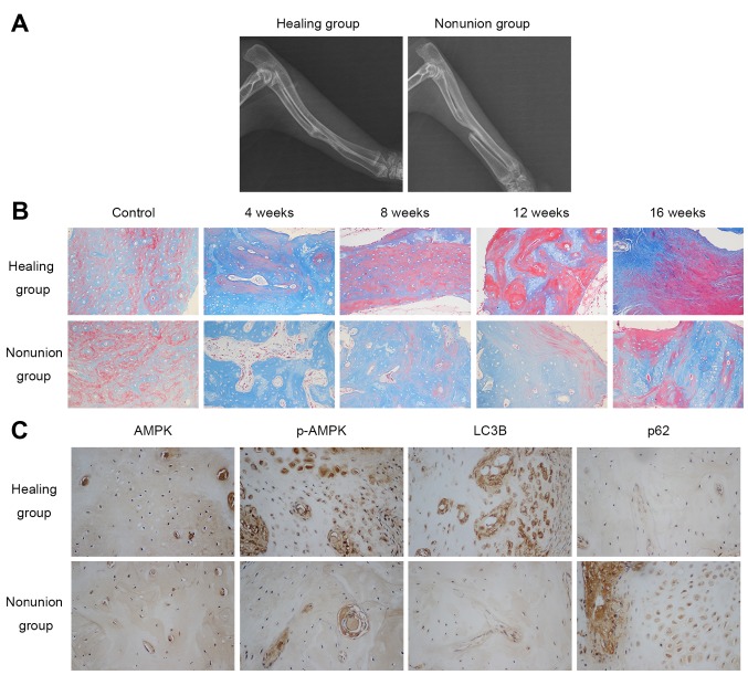
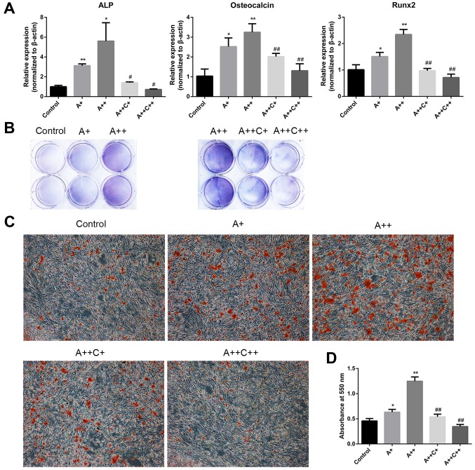
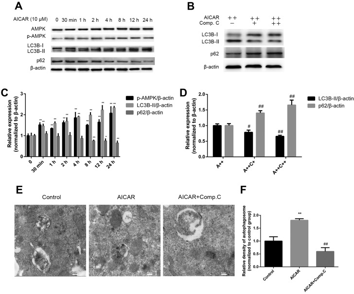
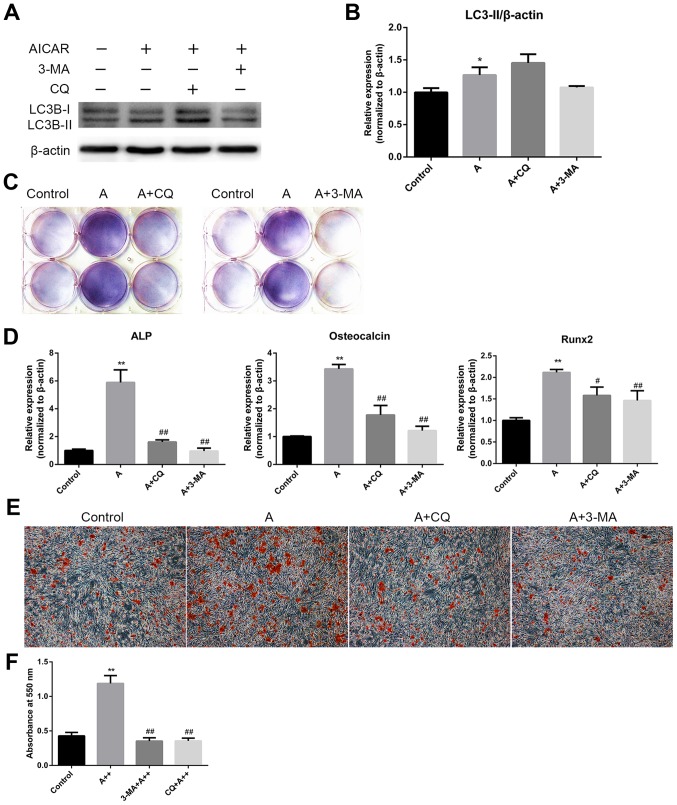
Previous studies have reported that adenosine monophosphate‑activated protein kinase (AMPK) activation can enhance osteoblast differentiation and mineralization; however, the underlying mechanism is not fully understood. Autophagy also serves an important role in osteoblast mineralization and bone homeostasis. The present study aimed to explore whether activation of AMPK could enhance osteoblast differentiation and mineralization via the induction of autophagy. The fracture healing and nonunion animal models were established and verified by X-ray imaging. Bone maturation was measured by Masson staining and the expression of AMPK, p-AMPK, microtubule-associated proteins 1A/1B light chain 3B II, and p62 in the fracture ends were detected by immunohistochemical staining. The mRNA expression levels of alkaline phosphatase (ALP), osteocalcin ,runt-related transcription factor 2 and BCN1 were determined by reverse transcription-quantitative polymerase chain reaction. 5-Bromo-4-chloro-3-indolyl phosphate/nitro blue tetrazolium staining was used to determine ALP activity and alizarin red staining was adopted to examine mineralization. Western blot analysis was performed to detect protein expression. Autophagosome was observed by Transmission electron microscopy. Small interfering (si)RNA was used to knock down the expression of target gene. In vivo experiments demonstrated that new bone mineralization and maturation was markedly restrained in the nonunion group, alongside decreased AMPK activation and autophagic activity, compared with in the fracture healing group. The results of an in vitro study indicated that AMPK activation stimulated the osteogenic differentiation of MC3T3‑E1 cells, with increases in ALP activity, mineralization, and the mRNA expression levels of ALP, osteocalcin and runt-related transcription factor 2. Furthermore, AMPK activation induced autophagy, as determined by upregulation of microtubule‑associated proteins 1A/1B light chain 3B, increased autophagosome density and downregulation of p62. In addition, inhibition of autophagy reversed the effects of AMPK activation on osteoblast differentiation. These results suggested that AMPK activation may stimulate osteoblast differentiation and mineralization via the induction of autophagy, and provides evidence to suggest that enhancing AMPK activation and autophagic activity may be a potential novel approach to promote fracture healing.
先前的研究报告称,一磷酸腺苷激活的蛋白激酶 (AMPK) 的激活可以增强成骨细胞分化和矿化;然而,其潜在机制尚不完全清楚。自噬在成骨细胞矿化和骨稳态中也起着重要作用。本研究旨在探讨 AMPK 的激活是否可以通过诱导自噬来增强成骨细胞分化和矿化。通过 X 射线成像建立并验证骨折愈合和非愈合动物模型。通过 Masson 染色测量骨成熟度,并通过免疫组织化学染色检测骨折端 AMPK、p-AMPK、微管相关蛋白 1A/1B 轻链 3B II 和 p62 的表达。通过逆转录-定量聚合酶链反应测定碱性磷酸酶 (ALP)、骨钙素、 runt 相关转录因子 2 和 BCN1 的 mRNA 表达水平。5-溴-4-氯-3-吲哚磷酸/硝基蓝四唑染色用于测定 ALP 活性,茜素红染色用于检测矿化。采用 Western blot 分析检测蛋白表达。通过透射电子显微镜观察自噬体。使用小干扰 (si)RNA 敲低靶基因的表达。体内实验表明,与骨折愈合组相比,非愈合组新骨矿化和成熟明显受到抑制,AMPK 激活和自噬活性降低。体外研究结果表明,AMPK 激活刺激 MC3T3-E1 细胞的成骨分化,ALP 活性、矿化以及 ALP、骨钙素和 runt 相关转录因子 2 的 mRNA 表达水平均增加。此外,AMPK 激活诱导自噬,表现为微管相关蛋白 1A/1B 轻链 3B 上调、自噬体密度增加和 p62 下调。此外,自噬抑制逆转了 AMPK 激活对成骨细胞分化的影响。这些结果表明,AMPK 激活可能通过诱导自噬来刺激成骨细胞分化和矿化,并为增强 AMPK 激活和自噬活性可能是促进骨折愈合的一种新方法提供了证据。