School of Medical Laboratory, Tianjin Medical University, Tianjin 300070, P.R. China.
Department of Clinical Laboratory Medicine, Taizhou University Hospital, Taizhou, Zhejiang 318000, P.R. China.
Int J Mol Med. 2019 Aug;44(2):652-660. doi: 10.3892/ijmm.2019.4216. Epub 2019 May 30.
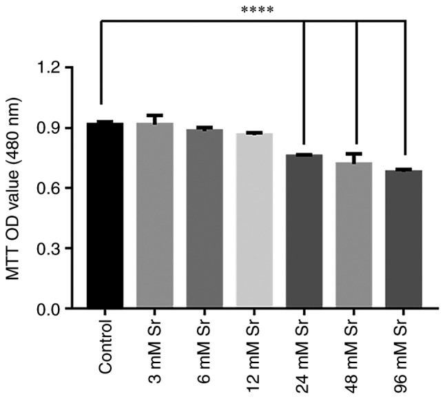
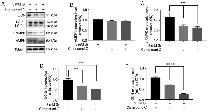
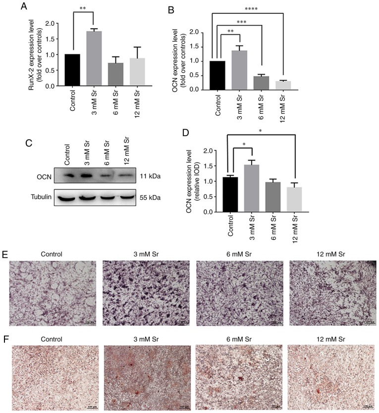
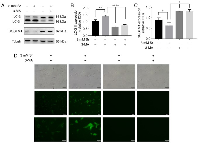
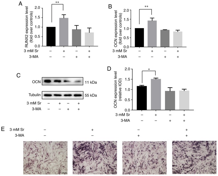
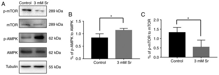
Strontium (Sr) is an alkaline earth metal that exerts the dual effect of improving bone formation and suppressing bone resorption, resulting in increased bone apposition rates and bone mineral density. However, the mechanisms through which Sr exerts these beneficial effects on bone have yet to be fully elucidated. The present study aimed to reveal the underlying molecular mechanisms associated with Sr‑induced osteogenic differentiation. The effects of Sr on cell proliferation and osteogenic differentiation were analyzed by MTT assay, RT‑qPCR, western blot analysis, alkaline phosphatase (ALP) and Alizarin red staining assays. The extent of autophagy was determined by monodansylcadaverine (MDC) staining and western blot analysis of two markers of cellular autophagic activity, the steatosis‑associated protein, sequestosome‑1 (SQSTM1/p62), and the two isoforms of microtubule‑associated protein 1 light chain 3 (LC3), LC‑3‑I/II. The expression levels of AMP‑activated protein kinase (AMPK) and mammalian target of rapamycin (mTOR) were also detected by western blot analysis. Sr at a concentration of 3 mM exerted the most pronounced effect on osteogenic differentiation, without any apparent cell toxicity. At the same time, cellular autophagy was active during this process. Subsequently, autophagy was blocked by 3‑methyladenine, and the enhancement of osteogenic differentiation in response to Sr was abrogated. Additionally, the phosphorylation level of AMPK was significantly increased, whereas that of mTOR was significantly decreased, in the Sr‑treated group. Taken together, the findings of the present study demonstrate that Sr stimulates AMPK‑activated autophagy to induce the osteogenic differentiation of MC3T3‑E1 cells.
锶(Sr)是一种碱土金属,具有促进骨形成和抑制骨吸收的双重作用,导致骨形成率和骨矿物质密度增加。然而,锶对骨骼产生这些有益作用的机制尚未完全阐明。本研究旨在揭示与锶诱导成骨分化相关的潜在分子机制。通过 MTT 检测、RT-qPCR、western blot 分析、碱性磷酸酶(ALP)和茜素红染色检测分析锶对细胞增殖和成骨分化的影响。通过单丹磺酰尸胺(MDC)染色和细胞自噬活性的两个标志物,即脂肪蓄积相关蛋白,sequestosome-1(SQSTM1/p62)和微管相关蛋白 1 轻链 3(LC3)的两种同工型,western blot 分析来确定自噬程度,LC3-I/II。通过 western blot 分析还检测了 AMP 激活的蛋白激酶(AMPK)和哺乳动物雷帕霉素靶蛋白(mTOR)的表达水平。浓度为 3 mM 的锶对成骨分化的作用最为明显,而没有明显的细胞毒性。同时,在此过程中细胞自噬活跃。随后,通过 3-甲基腺嘌呤阻断自噬,阻断了 Sr 诱导的成骨分化增强。此外,Sr 处理组 AMPK 的磷酸化水平显著增加,而 mTOR 的磷酸化水平显著降低。综上所述,本研究结果表明,Sr 刺激 AMPK 激活的自噬诱导 MC3T3-E1 细胞的成骨分化。