Kojima Daigo, Beppu Takaaki, Saura Hiroaki, Sato Yuichi, Fujiwara Shunrou, Ogasawara Kuniaki
Department of Neurosurgery, Iwate Medical University, 19-1 Uchimaru, Morioka 020-8505 Japan.
Radiol Case Rep. 2017 Oct 27;13(1):220-224. doi: 10.1016/j.radcr.2017.09.021. eCollection 2018 Feb.
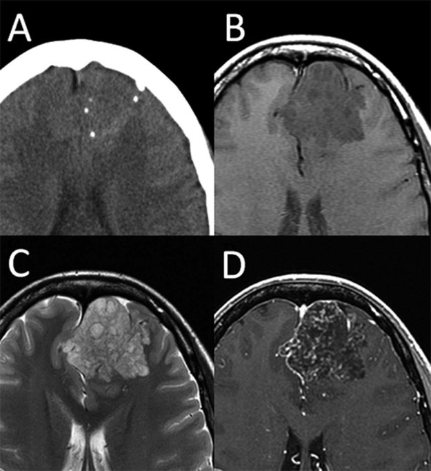
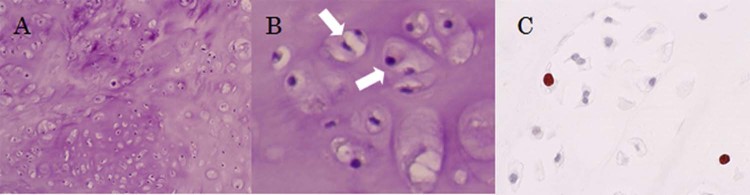
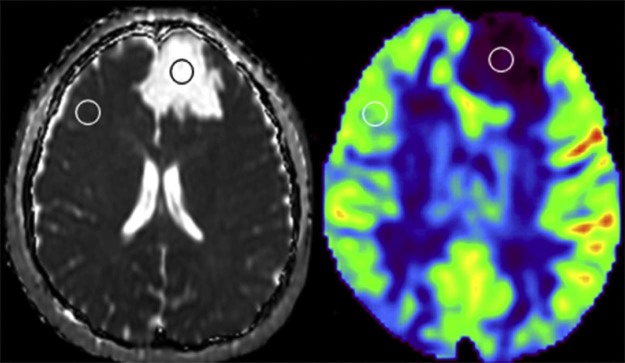
Intracranial chondrosarcoma is a very rare malignant tumor of the central nervous system, and is difficult to preoperatively distinguish from other tumors using conventional imaging techniques. Here, we report the case of a 24-year-old woman who presented with mild headache due to chondrosarcoma in the frontal lobe. Preoperative conventional images showed findings typical of an oligodendroglial tumor. However, high apparent diffusion coefficient (ADC) value and extreme hypoperfusion on arterial spin labeling (ASL) were inconsistent with oligodendroglial tumor characteristics. The tumor was completely removed using a standard surgical procedure. Histologic diagnosis was a conventional (classic) chondrosarcoma. High ADC and hypoperfusion on ASL represented low cellularity and low vascularity within conventional chondrosarcoma, respectively. We discuss the utility of ADC and ASL for the preoperative diagnosis of conventional chondrosarcoma.
颅内软骨肉瘤是一种非常罕见的中枢神经系统恶性肿瘤,术前使用传统成像技术很难将其与其他肿瘤区分开来。在此,我们报告一例24岁女性因额叶软骨肉瘤出现轻度头痛的病例。术前常规影像显示为少突胶质细胞瘤的典型表现。然而,高表观扩散系数(ADC)值和动脉自旋标记(ASL)上的极低灌注与少突胶质细胞瘤的特征不符。该肿瘤通过标准手术程序被完全切除。组织学诊断为传统型(经典型)软骨肉瘤。ASL上的高ADC值和低灌注分别代表传统软骨肉瘤内的低细胞密度和低血管密度。我们讨论了ADC和ASL在传统软骨肉瘤术前诊断中的应用价值。