Guo Yanxia, Walsh Alice M, Canavan Mary, Wechalekar Mihir D, Cole Suzanne, Yin Xuefeng, Scott Brittney, Loza Mathew, Orr Carl, McGarry Trudy, Bombardieri Michele, Humby Frances, Proudman Susanna M, Pitzalis Costantino, Smith Malcolm D, Friedman Joshua R, Anderson Ian, Madakamutil Loui, Veale Douglas J, Fearon Ursula, Nagpal Sunil
Immunology, Janssen Research, Pennsylvania, United States of America.
Molecular Rheumatology, Trinity Biomedical Sciences Institute, Trinity College, Dublin, Ireland.
PLoS One. 2018 Feb 28;13(2):e0192704. doi: 10.1371/journal.pone.0192704. eCollection 2018.
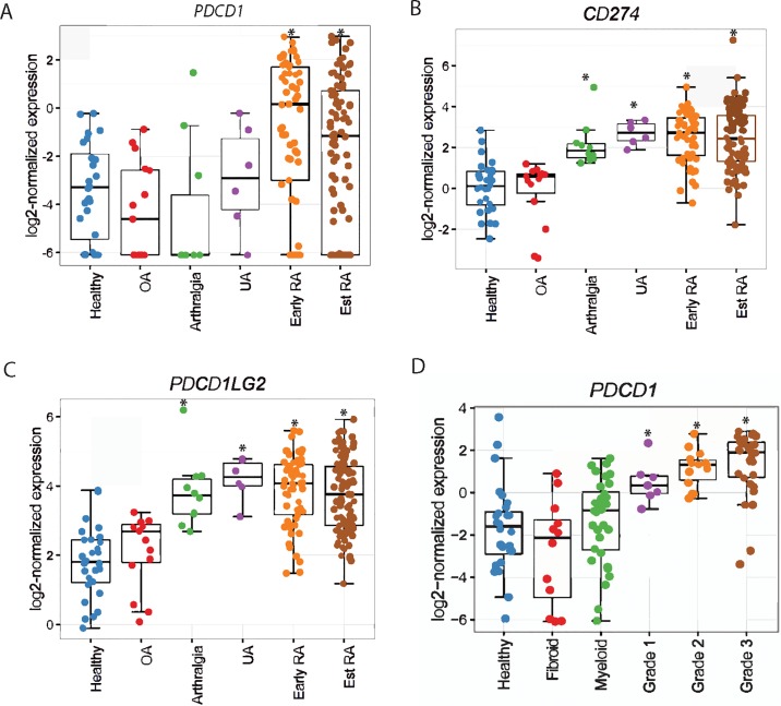
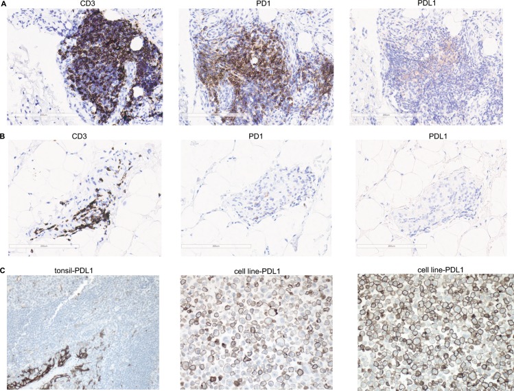
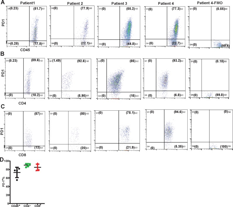
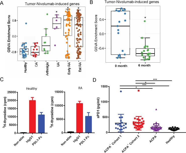
Immune checkpoint blockade with therapeutic anti-cytotoxic T lymphocyte-associated antigen (CTLA)-4 (Ipilimumab) and anti-programmed death (PD)-1 (Nivolumab and Pembrolizumab) antibodies alone or in combination has shown remarkable efficacy in multiple cancer types, concomitant with immune-related adverse events, including arthralgia and inflammatory arthritis (IA) in some patients. Herein, using Nivolumab (anti-PD-1 antagonist)-responsive genes along with transcriptomics of synovial tissue from multiple stages of rheumatoid arthritis (RA) disease progression, we have interrogated the activity status of PD-1 pathway during RA development. We demonstrate that the expression of PD-1 was increased in early and established RA synovial tissue compared to normal and OA synovium, whereas that of its ligands, programmed death ligand-1 (PD-L1) and PD-L2, was increased at all the stages of RA disease progression, namely arthralgia, IA/undifferentiated arthritis, early RA and established RA. Further, we show that RA patients expressed PD-1 on a majority of synovial tissue infiltrating CD4+ and CD8+ T cells. Moreover, enrichment of Nivolumab gene signature was observed in IA and RA, indicating that the PD-1 pathway was downregulated during RA disease progression. Furthermore, serum soluble (s) PD-1 levels were increased in autoantibody positive early RA patients. Interestingly, most of the early RA synovium tissue sections showed negative PD-L1 staining by immunohistochemistry. Therefore, downregulation in PD-1 inhibitory signaling in RA could be attributed to increased serum sPD-1 and decreased synovial tissue PD-L1 levels. Taken together, these data suggest that agonistic PD1 antibody-based therapeutics may show efficacy in RA treatment and interception.
单独或联合使用治疗性抗细胞毒性T淋巴细胞相关抗原(CTLA)-4(伊匹单抗)和抗程序性死亡(PD)-1(纳武单抗和帕博利珠单抗)抗体进行免疫检查点阻断,已在多种癌症类型中显示出显著疗效,同时伴有免疫相关不良事件,包括一些患者出现关节痛和炎性关节炎(IA)。在此,我们利用纳武单抗(抗PD-1拮抗剂)反应性基因以及类风湿关节炎(RA)疾病进展多个阶段滑膜组织的转录组学,探究了RA发展过程中PD-1通路的活性状态。我们证明,与正常和骨关节炎(OA)滑膜相比,PD-1在早期和确诊的RA滑膜组织中的表达增加,而其配体程序性死亡配体-1(PD-L1)和PD-L2的表达在RA疾病进展的所有阶段,即关节痛、IA/未分化关节炎、早期RA和确诊RA中均增加。此外,我们表明RA患者在大多数滑膜组织浸润的CD4+和CD8+T细胞上表达PD-1。而且,在IA和RA中观察到纳武单抗基因特征的富集,表明在RA疾病进展过程中PD-1通路被下调。此外,自身抗体阳性的早期RA患者血清可溶性(s)PD-1水平升高。有趣的是,大多数早期RA滑膜组织切片经免疫组织化学检测显示PD-L1染色为阴性。因此,RA中PD-1抑制信号的下调可能归因于血清sPD-1增加和滑膜组织PD-L1水平降低。综上所述,这些数据表明基于激动性PD1抗体的疗法可能在RA治疗和干预中显示出疗效。