Trampus Peter, Goepfert Christine, Welle Monika, Henke Diana, Forterre Franck, Schweizer-Gorgas Daniela
Division of Clinical Radiology, Vetsuisse-Faculty, University of Bern, Bern, Switzerland.
Institute of Animal Pathology, Vetsuisse-Faculty, University of Bern, Bern, Switzerland.
Front Vet Sci. 2018 Feb 15;5:16. doi: 10.3389/fvets.2018.00016. eCollection 2018.
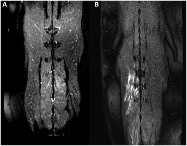
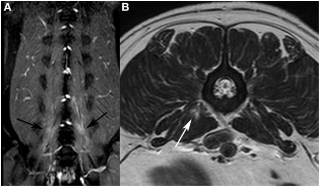
Muscle signal alteration detected on MRI is seen in diverse pathologic conditions. We observed signal alterations within the paraspinal muscles in dogs with acute thoracolumbar intervertebral disk extrusion. The aim of this retrospective study was to describe MRI features of paraspinal muscle signal alteration in dogs with acute thoracolumbar intervertebral disk extrusion and to investigate an association of the signal alterations with neurological grade, type and location of intervertebral disk extrusion, degree of spinal cord compression, and presence of epidural hemorrhage. Medical records of dogs undergoing MRI because of thoracolumbar intervertebral disk extrusion between August 2014 and June 2016 were reviewed. MRI was evaluated for SI changes within the paravertebral musculature, their location, extension, affected muscles, contrast enhancement, and signal void in T2* sequences. Intervertebral disk herniation was categorized as acute non-compressive nucleus pulposus extrusion (ANNPE) or compressive intervertebral disk disease. In five patients, muscle biopsies of areas with signal intensity changes were taken during surgery. In total, 103 dogs were enrolled in the study. Paraspinal muscle signal alterations were visible in 37 dogs (36%) affecting the epaxial musculature ( = 17), hypaxial musculature ( = 12), or both ( = 8). All signal alterations were hyperintense on T2-weighted images and iso- or hypointense in T1-weighted images. Signal void in T2* was not observed in any dog. Postcontrast sequences were available in 30 of the 37 dogs and showed enhancement in 45%. There was neither an association with degree of compression nor epidural hemorrhage. Intervertebral disk extrusion caudal to L1 and a higher neurological grade was associated with the presence of muscle changes. Histopathology revealed mild to moderate acute muscle fiber degeneration with edema and necrosis in three of five samples. The MRI, as well as the muscle samples, show rather unspecific changes. The underlying pathomechanism might be related to ischemia or muscle spasm, but also denervation edema may explain the signal alteration.
MRI检测到的肌肉信号改变可见于多种病理状况。我们在患有急性胸腰椎椎间盘突出的犬类中观察到椎旁肌肉内的信号改变。这项回顾性研究的目的是描述患有急性胸腰椎椎间盘突出的犬类椎旁肌肉信号改变的MRI特征,并研究信号改变与神经学分级、椎间盘突出的类型和位置、脊髓压迫程度以及硬膜外出血之间的关联。回顾了2014年8月至2016年6月期间因胸腰椎椎间盘突出接受MRI检查的犬类的病历。对MRI评估椎旁肌肉组织内的信号强度变化、其位置、范围、受影响的肌肉、对比增强以及T2序列中的信号缺失情况。椎间盘突出分为急性非压迫性髓核突出(ANNPE)或压迫性椎间盘疾病。在5例患者中,手术期间对信号强度改变区域进行了肌肉活检。总共103只犬纳入了该研究。37只犬(36%)可见椎旁肌肉信号改变,影响轴上肌肉(n = 17)、轴下肌肉(n = 12)或两者(n = 8)。所有信号改变在T2加权图像上呈高信号,在T1加权图像上呈等信号或低信号。未在任何犬中观察到T2信号缺失。37只犬中的30只可获得增强后序列,45%显示有增强。信号改变与压迫程度和硬膜外出血均无关联。L1以下的椎间盘突出和较高的神经学分级与肌肉改变的存在相关。组织病理学显示5个样本中的3个有轻度至中度急性肌纤维变性伴水肿和坏死。MRI以及肌肉样本显示出相当非特异性的改变。潜在的发病机制可能与缺血或肌肉痉挛有关,但失神经水肿也可能解释信号改变。