1 Department of Oncology, Cancer Research UK & Medical Research Council Oxford Institute for Radiation Oncology, University of Oxford, Oxford, UK.
2 Wellcome Centre for Integrative Neuroimaging, FMRIB Division, University of Oxford, John Radcliffe Hospital, Headington, Oxford, UK.
J Cereb Blood Flow Metab. 2019 Aug;39(8):1557-1569. doi: 10.1177/0271678X18756218. Epub 2018 Mar 2.
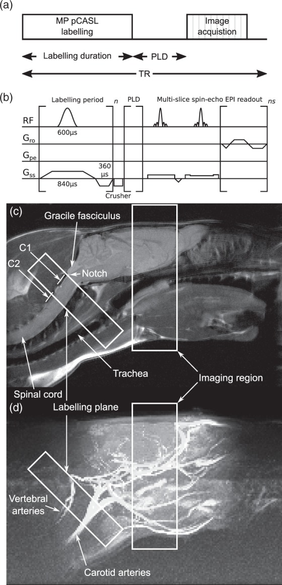
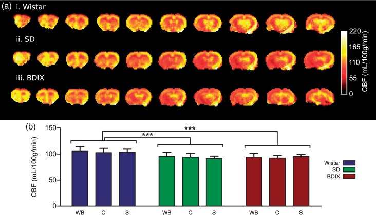
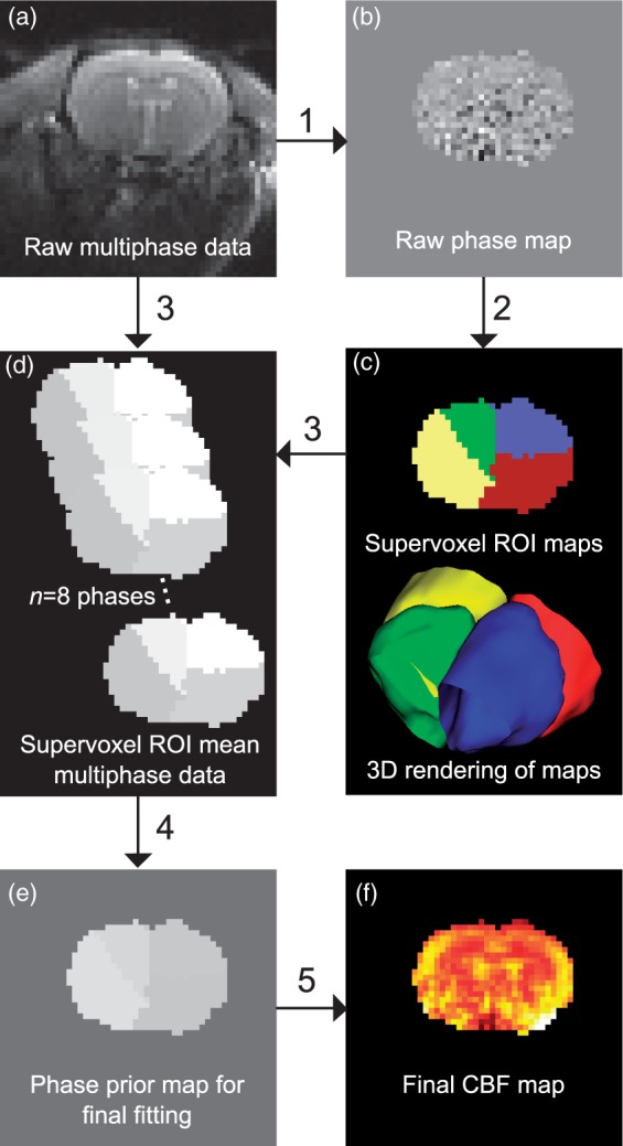
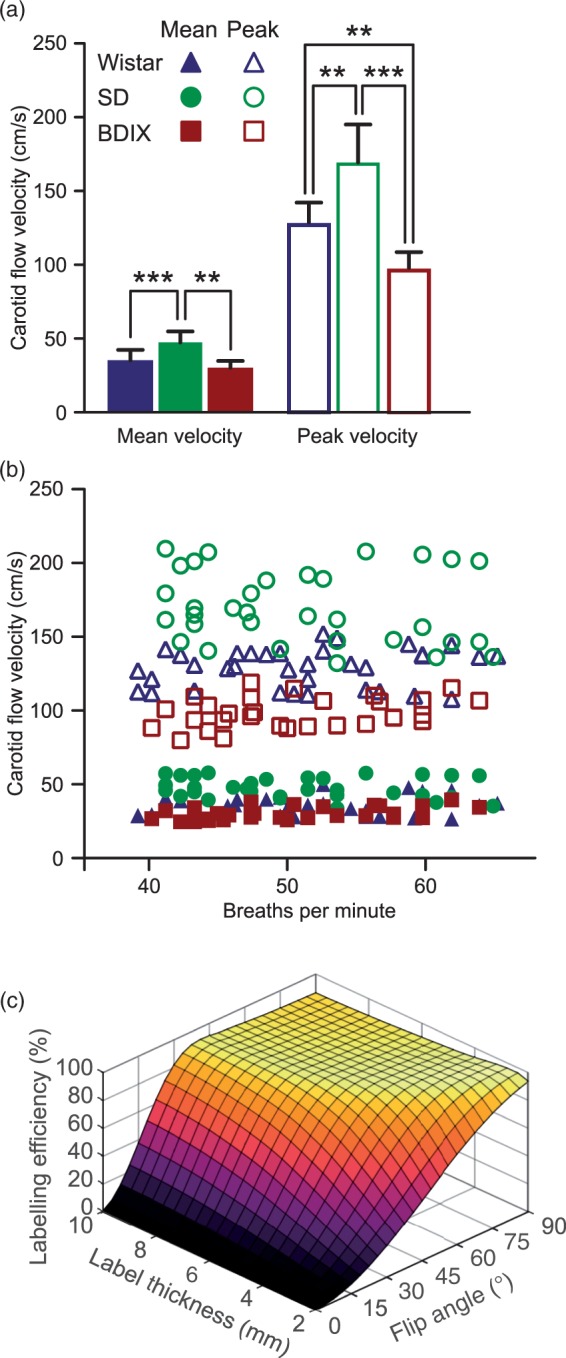
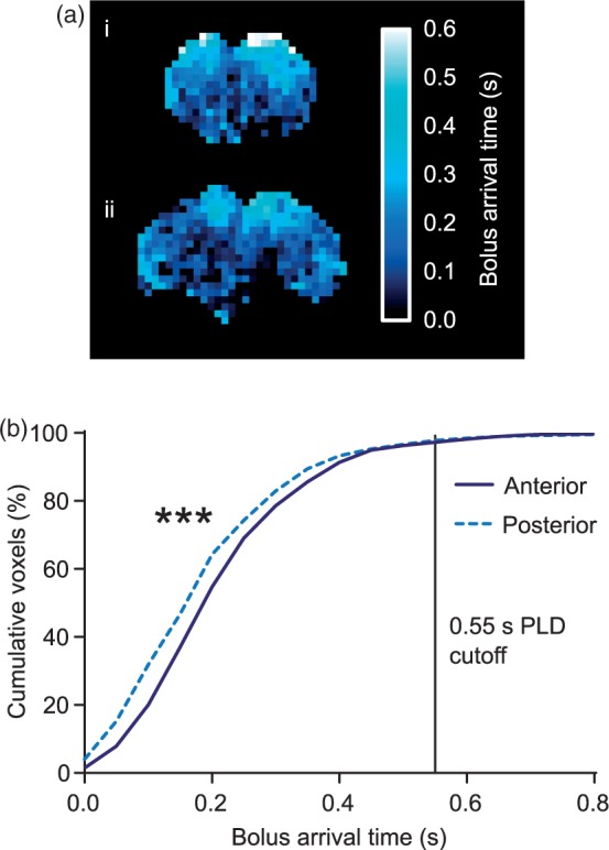
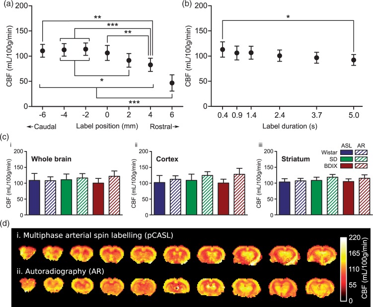
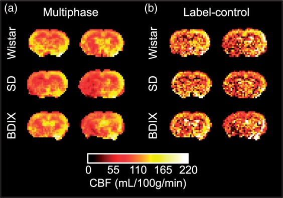
Cerebral blood flow is an important parameter in many diseases and functional studies that can be accurately measured in humans using arterial spin labelling (ASL) MRI. However, although rat models are frequently used for preclinical studies of both human disease and brain function, rat CBF measurements show poor consistency between studies. This lack of reproducibility is due, partly, to the smaller size and differing head geometry of rats compared to humans, as well as the differing analysis methodologies employed and higher field strengths used for preclinical MRI. To address these issues, we have implemented, optimised and validated a multiphase pseudo-continuous ASL technique, which overcomes many of the limitations of rat CBF measurement. Three rat strains (Wistar, Sprague Dawley and Berlin Druckrey IX) were used, and CBF values validated against gold-standard autoradiography measurements. Label positioning was found to be optimal at 45°, while post-label delay was optimised to 0.55 s. Whole brain CBF measures were 109 ± 22, 111 ± 18 and 100 ± 15 mL/100 g/min by multiphase pCASL, and 108 ± 12, 116 ± 14 and 122 ± 16 mL/100 g/min by autoradiography in Wistar, SD and BDIX cohorts, respectively. Tumour model analysis shows that the developed methods also apply in disease states. Thus, optimised multiphase pCASL provides robust, reproducible and non-invasive measurement of CBF in rats.
脑血流是许多疾病和功能研究中的一个重要参数,可以使用动脉自旋标记(ASL)MRI 对人类进行准确测量。然而,尽管大鼠模型经常被用于人类疾病和大脑功能的临床前研究,但大鼠 CBF 测量值在不同研究之间的一致性较差。这种可重复性的缺乏部分是由于大鼠与人类相比体积较小,头部几何形状不同,以及用于临床前 MRI 的分析方法和更高的场强不同。为了解决这些问题,我们已经实施、优化和验证了一种多相伪连续 ASL 技术,该技术克服了大鼠 CBF 测量的许多限制。使用了三种大鼠品系(Wistar、Sprague Dawley 和 Berlin Druckrey IX),并将 CBF 值与金标准放射性自显影测量值进行了验证。标签定位在 45°时被发现是最佳的,而延迟时间则被优化到 0.55s。通过多相 pCASL,全脑 CBF 测量值分别为 109±22、111±18 和 100±15mL/100g/min,而在 Wistar、SD 和 BDIX 组中,通过放射性自显影测量值分别为 108±12、116±14 和 122±16mL/100g/min。肿瘤模型分析表明,所开发的方法也适用于疾病状态。因此,优化的多相 pCASL 为大鼠提供了稳健、可重复和非侵入性的 CBF 测量。