Rodríguez-Razón Christian M, Yañez-Sánchez Irinea, Ramos-Santillan Vicente O, Velásquez-Ordóñez Celso, Gutiérrez-Rubio Susan A, García-García Maritza R, López-Roa Roció I, Sánchez-Hernández Pedro E, Daneri-Navarro Adrian, García-Iglesias Trinidad
Laboratory of Immunology and Institute of Experimental and Clinical Therapeutics, Department of Physiology, University Center of Health Sciences, University of Guadalajara, Jalisco, Mexico.
Center for Research in Nanosciences and Nanotechnology, Department of Natural and Exact Sciences, University Center of the Valleys, University of Guadalajara, Jalisco, Mexico.
Int J Nanomedicine. 2018 Feb 22;13:1081-1095. doi: 10.2147/IJN.S152237. eCollection 2018.
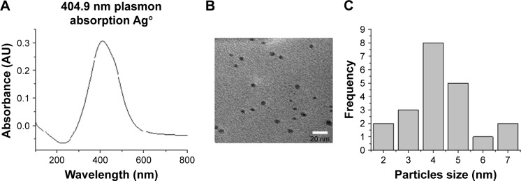
Silver nanoparticles (AgNPs) have attracted considerable attention due to the variety of their applications in medicine and other sciences. AgNPs have been used in vitro for treatment of various diseases, such as hepatitis B and herpes simplex infections as well as colon, cervical, and lung cancers. In this study, we assessed the effect on proliferation, adhesion, and apoptosis in breast cancer cell lines of different molecular profiles (MCF7, HCC1954, and HCC70) exposed to AgNPs (2-9 nm).
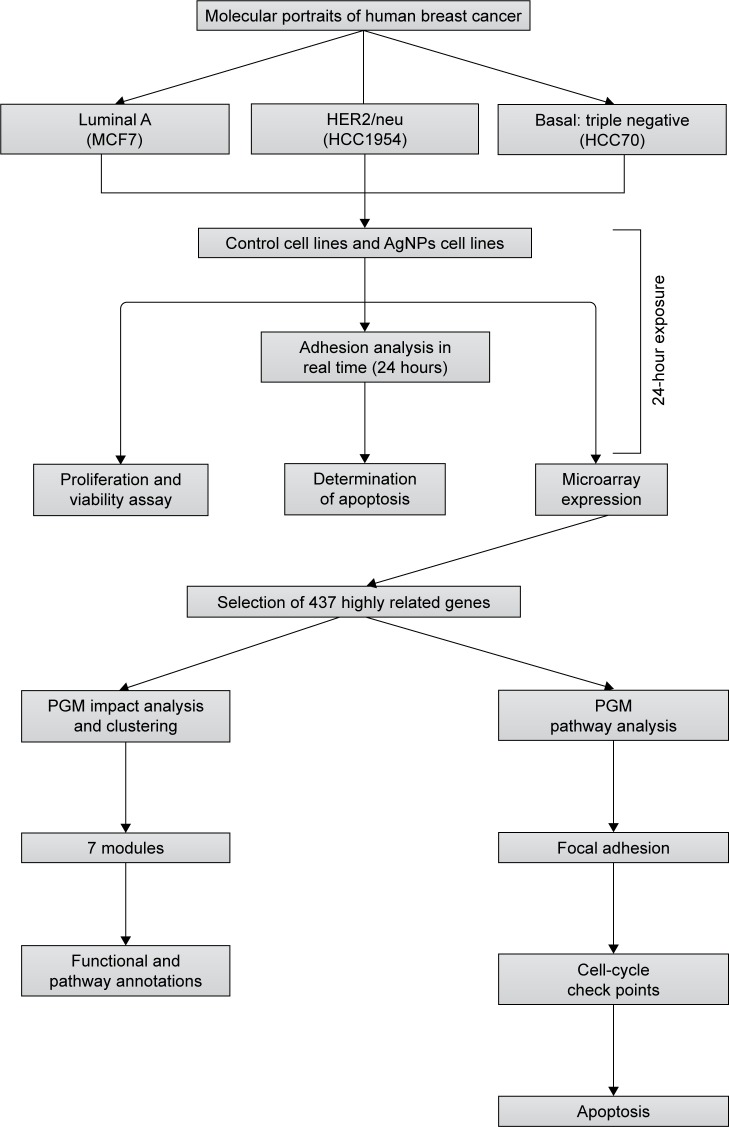
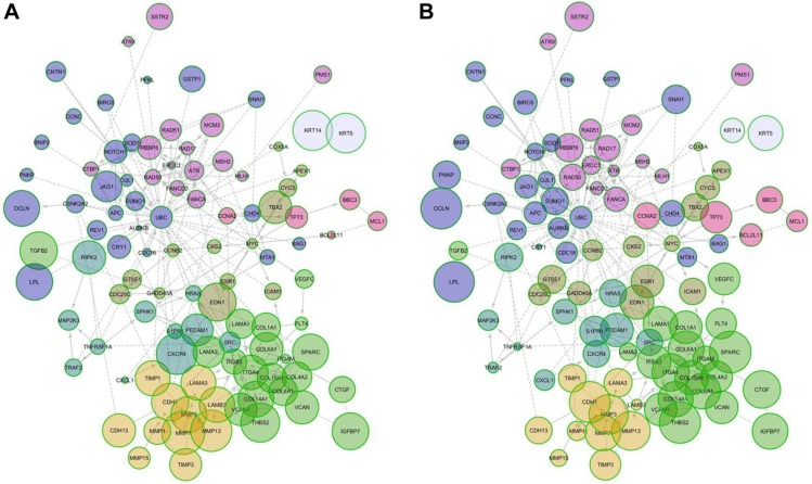
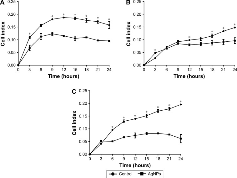
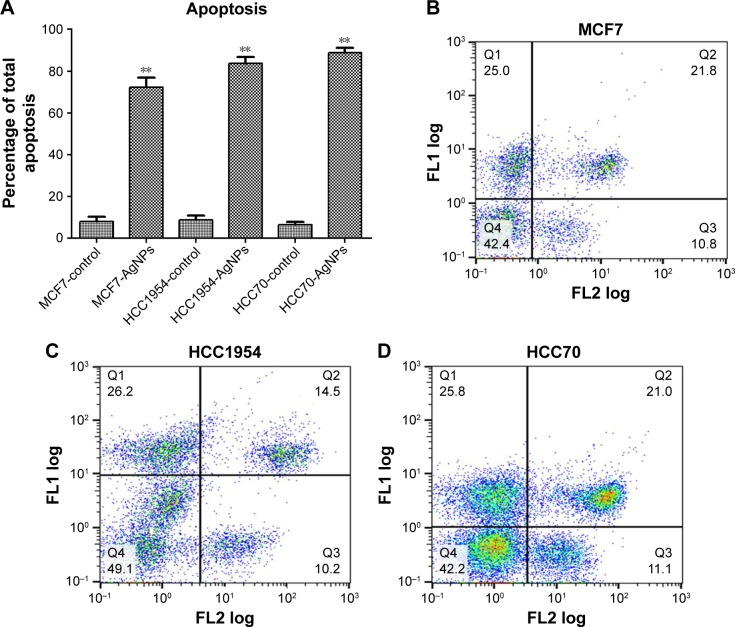
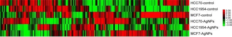
Breast cancer cell lines were incubated in vitro; MTT assay was used to assess proliferation. Adhesion was determined by real-time analysis with the xCELLingence system. Propidium iodide and fluorescein isothiocyanate-Annexin V assay were used to measure apoptosis. The transcriptome was assessed by gene expression microarray and Probabilistic Graphical Model (PGM) analyses.
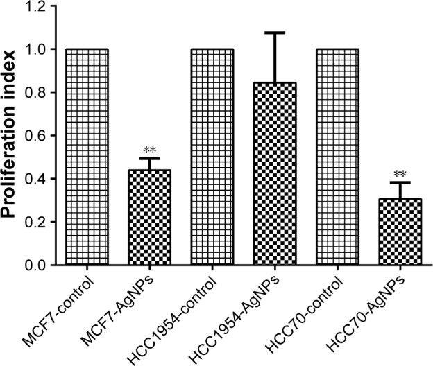
The results showed a decreased adhesion in breast cancer cell lines and the control exposed to AgNPs was noted in 24 hours (≤0.05). We observed a significant reduction in the proliferation of MCF7 and HCC70, but not in HCC1954. Apoptotic activity was seen in all cell lines exposed to AgNPs, with an apoptosis percentage of more than 60% in cancer cell lines and less than 60% in the control. PGM analysis confirmed, to some extent, the effects of AgNPs primarily on adhesion by changes in the extracellular matrix.
Exposure to AgNPs causes an antiproliferative, apoptotic, and anti-adhesive effect in breast cancer cell lines cultured in vitro. More research is needed to evaluate the potential use of AgNPs to treat different molecular profiles of breast cancer in humans.
银纳米颗粒(AgNPs)因其在医学和其他科学领域的多种应用而备受关注。AgNPs已在体外用于治疗各种疾病,如乙型肝炎、单纯疱疹感染以及结肠癌、宫颈癌和肺癌。在本研究中,我们评估了暴露于AgNPs(2 - 9纳米)对不同分子谱型(MCF7、HCC1954和HCC70)的乳腺癌细胞系增殖、黏附及凋亡的影响。
体外培养乳腺癌细胞系;采用MTT法评估增殖情况。使用xCELLigence系统通过实时分析确定黏附情况。采用碘化丙啶和异硫氰酸荧光素 - 膜联蛋白V法检测凋亡情况。通过基因表达微阵列和概率图形模型(PGM)分析评估转录组。
结果显示,暴露于AgNPs的乳腺癌细胞系和对照组在24小时时黏附能力下降(≤0.05)。我们观察到MCF7和HCC70的增殖显著降低,但HCC1954未出现此现象。在所有暴露于AgNPs的细胞系中均观察到凋亡活性,癌细胞系的凋亡率超过60%,对照组则低于60%。PGM分析在一定程度上证实了AgNPs主要通过细胞外基质的变化对黏附产生影响。
暴露于AgNPs会对体外培养的乳腺癌细胞系产生抗增殖、促凋亡和抗黏附作用。需要更多研究来评估AgNPs在治疗人类不同分子谱型乳腺癌方面的潜在用途。