• 文献检索
  • 文档翻译
  • 深度研究
  • 学术资讯
  • Suppr Zotero 插件Zotero 插件
  • 邀请有礼
  • 套餐&价格
  • 历史记录
应用&插件
Suppr Zotero 插件Zotero 插件浏览器插件Mac 客户端Windows 客户端微信小程序
定价
高级版会员购买积分包购买API积分包
服务
文献检索文档翻译深度研究API 文档MCP 服务
关于我们
关于 Suppr公司介绍联系我们用户协议隐私条款
关注我们

Suppr 超能文献

核心技术专利:CN118964589B侵权必究
粤ICP备2023148730 号-1Suppr @ 2026

文献检索

告别复杂PubMed语法,用中文像聊天一样搜索,搜遍4000万医学文献。AI智能推荐,让科研检索更轻松。

立即免费搜索

文件翻译

保留排版,准确专业,支持PDF/Word/PPT等文件格式,支持 12+语言互译。

免费翻译文档

深度研究

AI帮你快速写综述,25分钟生成高质量综述,智能提取关键信息,辅助科研写作。

立即免费体验

与1型特发性黄斑毛细血管扩张症视力相关的预测性多影像生物标志物。

Predictive multi-imaging biomarkers relevant for visual acuity in idiopathic macular telangiectasis type 1.

作者信息

Guo Jingli, Tang WenYi, Ye Xiaofeng, Wu Haixiang, Xu Gezhi, Liu Wei, Zhang Yongjin

机构信息

Department of Ophthalmology, Eye and ENT Hospital of Fudan University, Shanghai Key Laboratory of Visual Impairment and Restoration, Shanghai, 200031, China.

出版信息

BMC Ophthalmol. 2018 Mar 5;18(1):69. doi: 10.1186/s12886-018-0737-y.

DOI:10.1186/s12886-018-0737-y
PMID:29506510
原文链接:https://pmc.ncbi.nlm.nih.gov/articles/PMC5836363/
Abstract

BACKGROUND

To evaluate the structural changes associated with visual acuity (VA) in patients with idiopathic macular telangiectasia (MT) type 1 using multimodal imaging modalities.

METHODS

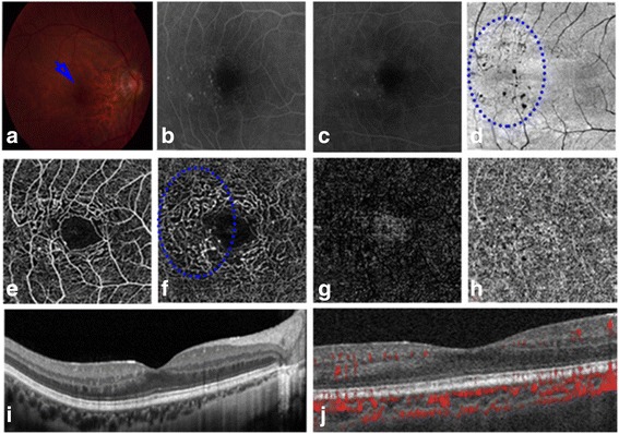
A retrospective study of 14 patients with MT type 1 and of 10 eyes from 10 healthy individuals as age-matched controls was conducted. The medical records of patients who had undergone colour fundus photography, spectral domain optical coherence tomography (OCT), fluorescein angiography and OCT angiography were reviewed. Central macular thickness (CMT), the areas of macular oedema and ellipsoid zone (EZ) disruption, EZ length, disorganization of the retinal inner layers (DRIL) and external limiting membrane (ELM) disruption, as measured by spectral domain OCT; and vascular density and the foveal avascular zones (FAZ) of the superficial capillary plexus (SCP) and deep capillary plexus (DCP), as measured by OCT angiography, were assessed in MT type 1 eyes and correlated with VA.

RESULTS

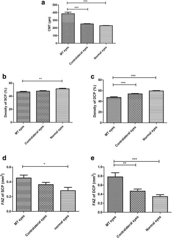
The mean baseline best-corrected VA of MT type 1 eyes was 0.45 ± 0.28. The mean CMT was 385.19 ± 75.21 μm in MT type 1 eyes and 252.43 ± 15.77 μm in contralateral eyes (Z = - 4.113, p < 0.001). The mean vessel density of the DCP was lower in MT type 1 eyes (47.25 ± 4.69%) than in contralateral eyes (53.93 ± 2.94%) and normal eyes (59.37 ± 2.50%) (Z = - 3.492, - 4.099; p < 0.001, < 0.001). The baseline logMAR VA was correlated with CMT (r = 0.682, p = 0.007), SCP density (r = - 0.652, p = 0.012), DCP density (r = - 0.700, p = 0.005), total area of EZ disruption (r = 0.649, p = 0.012); and total lengths of EZ (r = 0.681, p = 0.007), ELM (r = 0.699, p = 0.005) and DRIL (r = 0.707, p = 0.005) disruption in the 1-mm-diameter foveal region in MT type 1 eyes.

CONCLUSIONS

Decreased DCP density and the presence of DRIL may be predictive biomarkers of VA in MT type 1. CMT, SCP density, total area of EZ disruption, and lengths of EZ and ELM disruption within the 1-mm-diameter central region were strongly associated with VA.

摘要

背景

使用多模态成像方式评估1型特发性黄斑毛细血管扩张症(MT)患者与视力(VA)相关的结构变化。

方法

对14例1型MT患者以及10名健康个体的10只眼睛作为年龄匹配对照进行回顾性研究。回顾了接受彩色眼底照相、光谱域光学相干断层扫描(OCT)、荧光素血管造影和OCT血管造影的患者的病历。通过光谱域OCT测量中心黄斑厚度(CMT)、黄斑水肿面积和椭圆体带(EZ)破坏、EZ长度、视网膜内层紊乱(DRIL)和外界膜(ELM)破坏;通过OCT血管造影测量1型MT眼睛的血管密度以及浅表毛细血管丛(SCP)和深部毛细血管丛(DCP)的黄斑无血管区(FAZ),并将其与VA进行相关性分析。

结果

1型MT眼睛的平均基线最佳矫正视力为0.45±0.28。1型MT眼睛的平均CMT为385.19±75.21μm,对侧眼睛为252.43±15.77μm(Z = -4.113,p < 0.001)。1型MT眼睛的DCP平均血管密度(47.25±4.69%)低于对侧眼睛(53.93±2.94%)和正常眼睛(59.37±2.50%)(Z = -3.492,-4.099;p < 0.001,< 0.001)。基线logMAR视力与CMT(r = 0.682,p = 0.007)、SCP密度(r = -0.652,p = 0.012)、DCP密度(r = -0.700,p = 0.005)、EZ破坏总面积(r = 0.649,p = 0.012);以及1型MT眼睛直径1mm的黄斑区EZ总长度(r = 0.68l,p = 0.007)、ELM(r = 0.699,p = 0.005)和DRIL(r = 0.707,p = 0.005)破坏相关。

结论

DCP密度降低和DRIL的存在可能是1型MT患者VA的预测生物标志物。CMT、SCP密度、EZ破坏总面积以及直径1mm中心区域内的EZ和ELM破坏长度与VA密切相关。

https://cdn.ncbi.nlm.nih.gov/pmc/blobs/6b0c/5836363/455419e47ca0/12886_2018_737_Fig2_HTML.jpg
https://cdn.ncbi.nlm.nih.gov/pmc/blobs/6b0c/5836363/44dd38a6c006/12886_2018_737_Fig1_HTML.jpg
https://cdn.ncbi.nlm.nih.gov/pmc/blobs/6b0c/5836363/455419e47ca0/12886_2018_737_Fig2_HTML.jpg
https://cdn.ncbi.nlm.nih.gov/pmc/blobs/6b0c/5836363/44dd38a6c006/12886_2018_737_Fig1_HTML.jpg
https://cdn.ncbi.nlm.nih.gov/pmc/blobs/6b0c/5836363/455419e47ca0/12886_2018_737_Fig2_HTML.jpg

相似文献

1
Predictive multi-imaging biomarkers relevant for visual acuity in idiopathic macular telangiectasis type 1.与1型特发性黄斑毛细血管扩张症视力相关的预测性多影像生物标志物。
BMC Ophthalmol. 2018 Mar 5;18(1):69. doi: 10.1186/s12886-018-0737-y.
2
OCT Angiography and Ellipsoid Zone Mapping of Macular Telangiectasia Type 2 From the AVATAR Study.来自AVATAR研究的2型黄斑毛细血管扩张症的光学相干断层扫描血管造影及椭圆体带成像
Invest Ophthalmol Vis Sci. 2017 Jul 1;58(9):3683-3689. doi: 10.1167/iovs.16-20976.
3
[Correlation of capillary plexus with visual acuity in idiopathic macular epiretinal membrane eyes using optical coherence tomography angiography].[利用光学相干断层扫描血管造影术研究特发性黄斑视网膜前膜眼中毛细血管丛与视力的相关性]
Zhonghua Yan Ke Za Zhi. 2019 Oct 11;55(10):757-762. doi: 10.3760/cma.j.issn.0412-4081.2019.10.006.
4
Radiation Maculopathy After Proton Beam Therapy for Uveal Melanoma: Optical Coherence Tomography Angiography Alterations Influencing Visual Acuity.葡萄膜黑色素瘤质子束治疗后的放射性黄斑病变:影响视力的光学相干断层扫描血管造影改变
Invest Ophthalmol Vis Sci. 2017 Aug 1;58(10):3851-3861. doi: 10.1167/iovs.17-22324.
5
Disorganization of Retinal Inner Layers and Ellipsoid Zone Disruption Predict Visual Outcomes in Central Retinal Vein Occlusion.视网膜内层结构紊乱和椭圆体带破坏可预测视网膜中央静脉阻塞的视觉预后。
Ophthalmol Retina. 2019 Jan;3(1):83-92. doi: 10.1016/j.oret.2018.07.008. Epub 2018 Oct 19.
6
Association of Disorganization of Retinal Inner Layers With Visual Acuity In Eyes With Uveitic Cystoid Macular Edema.葡萄膜炎性黄斑囊样水肿患者视网膜内层结构紊乱与视力的相关性
Am J Ophthalmol. 2017 May;177:116-125. doi: 10.1016/j.ajo.2017.02.017. Epub 2017 Feb 22.
7
Evaluation of vessel density in disorganization of retinal inner layers after resolved diabetic macular edema using optical coherence tomography angiography.应用光相干断层扫描血管造影评估糖尿病黄斑水肿消退后视网膜内层组织紊乱的血管密度。
PLoS One. 2021 Jan 12;16(1):e0244789. doi: 10.1371/journal.pone.0244789. eCollection 2021.
8
Disorganization of the retinal inner layers as a predictor of visual acuity in eyes with center-involved diabetic macular edema.视网膜内层结构紊乱作为累及黄斑中心的糖尿病性黄斑水肿患眼视力的预测指标。
JAMA Ophthalmol. 2014 Nov;132(11):1309-16. doi: 10.1001/jamaophthalmol.2014.2350.
9
Factors based on optical coherence tomography correlated with vision impairment in diabetic patients.基于光学相干断层扫描的因素与糖尿病患者视力障碍的相关性。
Sci Rep. 2021 Feb 4;11(1):3004. doi: 10.1038/s41598-021-82334-w.
10
Assessment of macular microvasculature features before and after vitrectomy in the idiopathic macular epiretinal membrane using a grading system: An optical coherence tomography angiography study.采用分级系统评估特发性黄斑视网膜前膜玻璃体切除术前、后黄斑微血管特征:光学相干断层扫描血管造影研究。
Acta Ophthalmol. 2021 Nov;99(7):e1168-e1175. doi: 10.1111/aos.14753. Epub 2021 Jan 10.

引用本文的文献

1
Automated assessment of the smoothness of retinal layers in optical coherence tomography images using a machine learning algorithm.使用机器学习算法自动评估光学相干断层扫描图像中视网膜层的平滑度。
BMC Med Imaging. 2023 Feb 2;23(1):21. doi: 10.1186/s12880-023-00976-w.
2
Visualization of microaneurysms in macular telangiectasia type 1 on optical coherence tomography angiography before and after photocoagulation.在光相干断层扫描血管造影术下观察 1 型黄斑毛细血管扩张症在光凝前后的微动脉瘤。
Graefes Arch Clin Exp Ophthalmol. 2021 Jun;259(6):1513-1520. doi: 10.1007/s00417-020-04953-9. Epub 2020 Oct 2.
3
Incomplete Retinal Pigment Epithelial and Outer Retinal Atrophy in Age-Related Macular Degeneration: Classification of Atrophy Meeting Report 4.

本文引用的文献

1
Fluorescein Angiography Versus Optical Coherence Tomography Angiography in Macular Telangiectasia Type I Treated With Bevacizumab Therapy.在接受贝伐单抗治疗的Ⅰ型黄斑毛细血管扩张症中,荧光素血管造影与光学相干断层扫描血管造影的对比研究
Ophthalmic Surg Lasers Imaging Retina. 2017 Mar 1;48(3):263-266. doi: 10.3928/23258160-20170301-12.
2
Optical coherence tomographic angiography in type 1 idiopathic macular telangiectasia.1型特发性黄斑毛细血管扩张症的光学相干断层扫描血管造影
Clin Exp Optom. 2018 Jan;101(1):143-144. doi: 10.1111/cxo.12536. Epub 2017 Mar 14.
3
Association of Disorganization of Retinal Inner Layers With Visual Acuity In Eyes With Uveitic Cystoid Macular Edema.
年龄相关性黄斑变性的不完全性视网膜色素上皮和外层视网膜萎缩:萎缩分类会议报告 4。
Ophthalmology. 2020 Mar;127(3):394-409. doi: 10.1016/j.ophtha.2019.09.035. Epub 2019 Sep 30.
4
Disorganization of the retinal inner layers as a prognostic factor in eyes with central retinal artery occlusion.视网膜内层结构紊乱作为视网膜中央动脉阻塞患者眼睛的一个预后因素。
Int J Ophthalmol. 2019 Jun 18;12(6):990-995. doi: 10.18240/ijo.2019.06.18. eCollection 2019.
5
[Foveal avascular zone and OCT angiography. An overview of current knowledge].[黄斑无血管区与光学相干断层扫描血管造影。当前知识概述]
Ophthalmologe. 2019 Jul;116(7):610-616. doi: 10.1007/s00347-018-0838-2.
葡萄膜炎性黄斑囊样水肿患者视网膜内层结构紊乱与视力的相关性
Am J Ophthalmol. 2017 May;177:116-125. doi: 10.1016/j.ajo.2017.02.017. Epub 2017 Feb 22.
4
Optical coherence tomography angiography of macular telangiectasia type 1: Comparison with mild diabetic macular edema.1型黄斑毛细血管扩张症的光学相干断层扫描血管造影:与轻度糖尿病性黄斑水肿的比较。
Lasers Surg Med. 2017 Mar;49(3):225-232. doi: 10.1002/lsm.22645. Epub 2017 Feb 7.
5
Macular Vascular Flow Area and Vascular Density in Healthy Population Using Optical Coherence Tomography Angiography.使用光学相干断层扫描血管造影术测量健康人群的黄斑血管血流面积和血管密度
Invest Ophthalmol Vis Sci. 2016 Dec 1;57(15):6713. doi: 10.1167/iovs.16-20359.
6
Comparison of diabetic retinopathy classification using fluorescein angiography and optical coherence tomography angiography.使用荧光素血管造影术和光学相干断层扫描血管造影术对糖尿病视网膜病变分类的比较。
Br J Ophthalmol. 2017 Jan;101(1):62-68. doi: 10.1136/bjophthalmol-2016-309424. Epub 2016 Dec 7.
7
Characteristics of Retinal Neovascularization in Proliferative Diabetic Retinopathy Imaged by Optical Coherence Tomography Angiography.光学相干断层扫描血管造影成像的增殖性糖尿病视网膜病变中视网膜新生血管的特征
Invest Ophthalmol Vis Sci. 2016 Nov 1;57(14):6247-6255. doi: 10.1167/iovs.16-20210.
8
CORRELATION OF MICROVASCULAR STRUCTURES ON OPTICAL COHERENCE TOMOGRAPHY ANGIOGRAPHY WITH VISUAL ACUITY IN RETINAL VEIN OCCLUSION.视网膜静脉阻塞患者光学相干断层扫描血管造影微血管结构与视力的相关性
Retina. 2017 Sep;37(9):1700-1709. doi: 10.1097/IAE.0000000000001403.
9
Optical coherence angiography: A review.光学相干血管造影术:综述
Medicine (Baltimore). 2016 Oct;95(41):e4907. doi: 10.1097/MD.0000000000004907.
10
Visual Acuity Is Correlated with the Area of the Foveal Avascular Zone in Diabetic Retinopathy and Retinal Vein Occlusion.视力与糖尿病视网膜病变和视网膜静脉阻塞的中心无血管区面积相关。
Ophthalmology. 2016 Nov;123(11):2352-2367. doi: 10.1016/j.ophtha.2016.07.008. Epub 2016 Aug 11.