State Key Laboratory of Molecular Vaccinology and Molecular Diagnostics & Center for Molecular Imaging and Translational Medicine, School of Public Health, Xiamen University, Xiamen 361102, China.
Department of Nuclear Medicine, Zhongshan Hospital affiliated to Xiamen University, Xiamen 361004, Fujian, China.
Theranostics. 2018 Feb 2;8(5):1340-1349. doi: 10.7150/thno.22806. eCollection 2018.
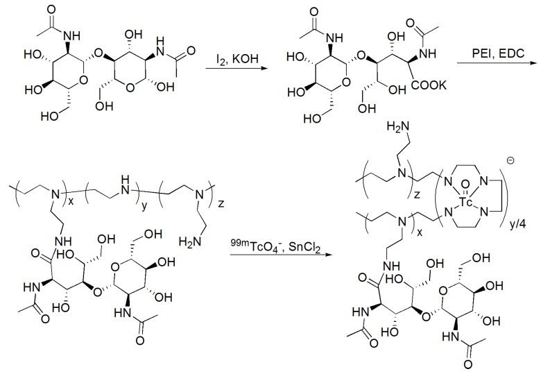
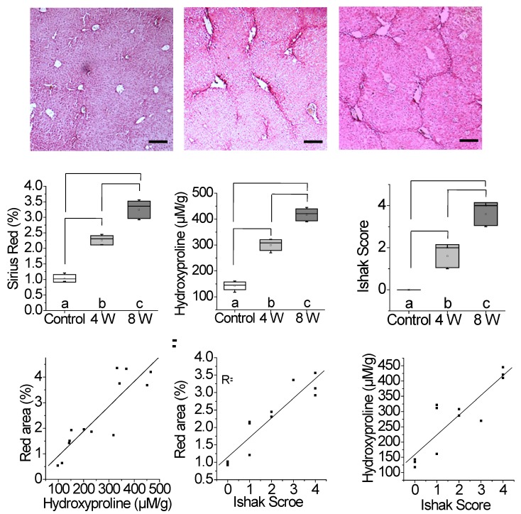
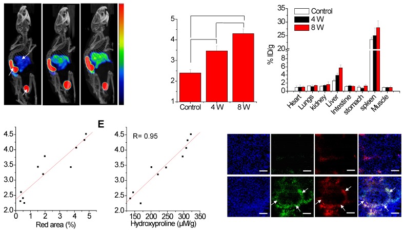
Extracellular matrix (ECM) accumulation in liver fibrosis is caused by the activation of hepatic stellate cells (HSCs). The goal of this study was to develop a Tc-labeled N-acetylglucosamine (GlcNAc) that specifically interacts with desmin and vimentin expressed on activated HSCs to monitor the progression and prognosis of liver fibrosis using single-photon emission computed tomography (SPECT) imaging. GlcNAc-conjugated polyethylenimine (PEI) was first prepared and radiolabeled with Tc. Noninvasive SPECT imaging with Tc-GlcNAc-PEI was used to assess liver fibrosis in a carbon tetrachloride (CCl) mouse model. The liver uptake value (LUV) of Tc-GlcNAc-PEI was measured by drawing the region of interest (ROI) of the whole liver as previously suggested. The LUV of the CCl groups was compared with that of the olive oil group. Next, we estimated the correlation between the results of SPECT imaging and physiological indexes. After treatment with clodronate liposome, the LUV of Tc-GlcNAc-PEI in fibrotic mice was compared with that in control mice. Tc-GlcNAc-PEI is a hydrophilic compound with high radiochemical purity (>98%) and good stability. It could specifically target desmin and vimentin on the surface of activated HSCs with high affinity (the K values were 53.75 ± 9.50 nM and 20.98 ± 3.56 nM, respectively). The LUV of Tc-GlcNAc-PEI was significantly different between the CCl and control groups as early as 4 weeks of CCl administration (3.30 ± 0.160 vs 2.34 ± 0.114%/cc; ˂ 0.05). There was a strong correlation between the LUV and Sirius Red quantification (R = 0.92, ˂ 0.001). Compared with control, clodronate liposome treatment reduced the LUV of Tc-GlcNAc-PEI (4.62 ± 0.352 vs 2.133 ± 0.414%/cc; ˂ 0.05). Tc-GlcNAc-PEI SPECT/CT was useful in assessing liver fibrosis and monitoring the treatment response.
细胞外基质(ECM)在肝纤维化中的积累是由肝星状细胞(HSCs)的激活引起的。本研究的目的是开发一种 Tc 标记的 N-乙酰葡萄糖胺(GlcNAc),它可以特异性地与活化的 HSCs 上表达的结蛋白和波形蛋白相互作用,使用单光子发射计算机断层扫描(SPECT)成像来监测肝纤维化的进展和预后。首先制备 GlcNAc 修饰的聚亚乙基亚胺(PEI),并用 Tc 标记。使用 Tc-GlcNAc-PEI 进行非侵入性 SPECT 成像,以评估四氯化碳(CCl)小鼠模型中的肝纤维化。如前所述,通过绘制整个肝脏的感兴趣区域(ROI)来测量 Tc-GlcNAc-PEI 的肝脏摄取值(LUV)。比较 CCl 组和橄榄油组的 LUV。接下来,我们估计 SPECT 成像结果与生理指标之间的相关性。在用氯膦酸盐脂质体治疗后,比较纤维化小鼠和对照组中 Tc-GlcNAc-PEI 的 LUV。Tc-GlcNAc-PEI 是一种亲水性化合物,具有高放射化学纯度(>98%)和良好的稳定性。它可以特异性地与活化的 HSCs 表面的结蛋白和波形蛋白高亲和力结合(K 值分别为 53.75±9.50 nM 和 20.98±3.56 nM)。早在给予 CCl 后 4 周,Tc-GlcNAc-PEI 的 LUV 在 CCl 组和对照组之间就有显著差异(3.30±0.160 与 2.34±0.114%/cc; ˂0.05)。LUV 与天狼星红定量之间存在很强的相关性(R=0.92, ˂0.001)。与对照组相比,氯膦酸盐脂质体治疗降低了 Tc-GlcNAc-PEI 的 LUV(4.62±0.352 与 2.133±0.414%/cc; ˂0.05)。Tc-GlcNAc-PEI SPECT/CT 可用于评估肝纤维化和监测治疗反应。