Huang Xiangsheng, Wiehr Stefan, Wild Anna-Maria, Voßberg Patrick, Hoffmann Wolfgang, Grüner Beate, Köhler Carsten, Soboslay Peter T
Institute for Tropical Medicine, Eberhard Karls University, Tübingen, Germany.
Werner Siemens Imaging Center, Department of Preclinical Imaging and Radiopharmacy, Eberhard Karls University, Tübingen, Germany.
Oncotarget. 2018 Jan 10;9(10):9073-9087. doi: 10.18632/oncotarget.24142. eCollection 2018 Feb 6.
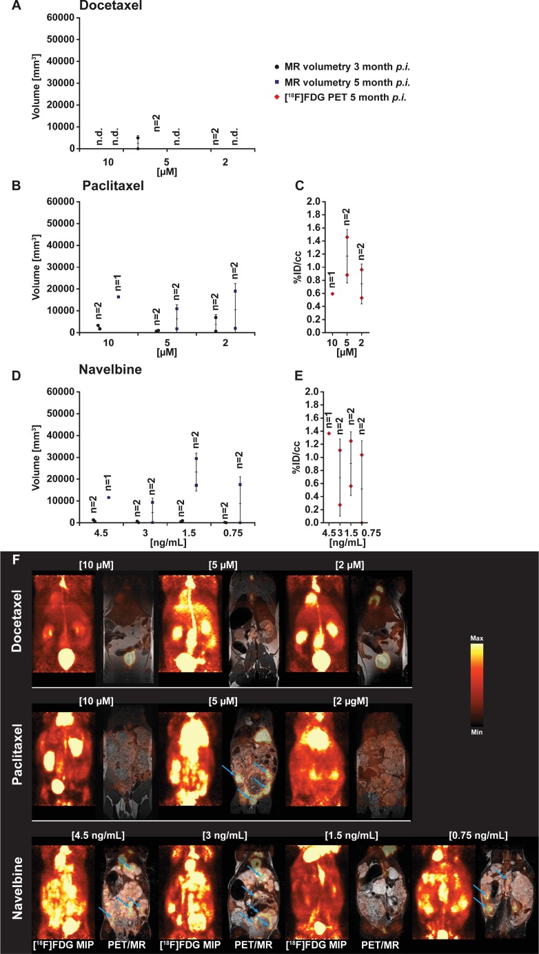
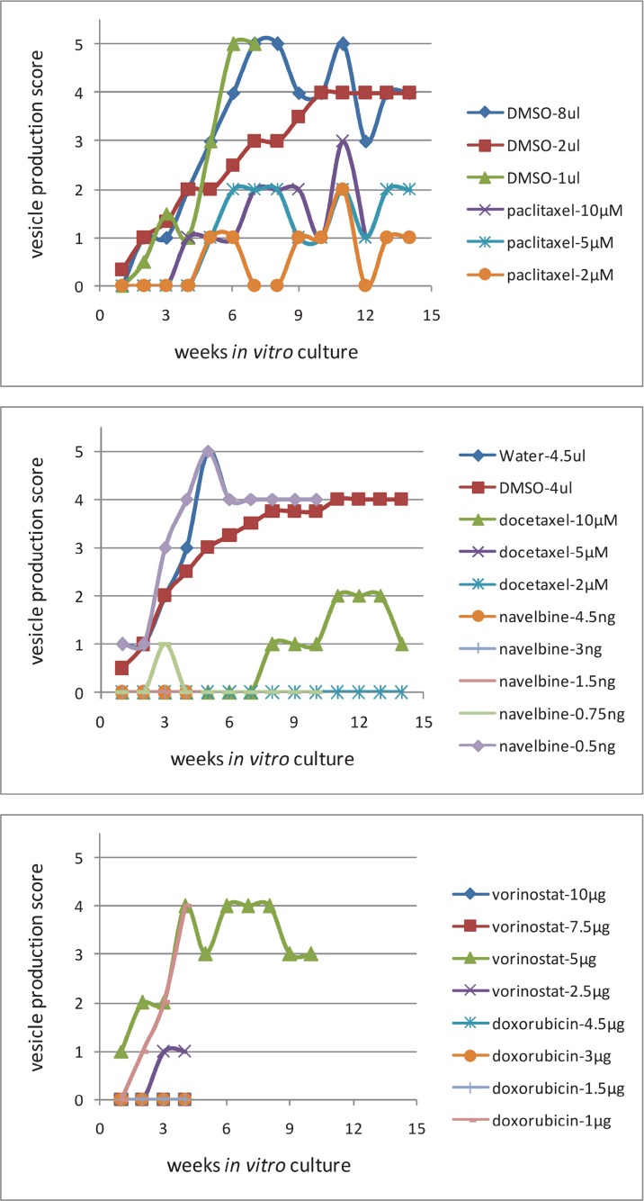
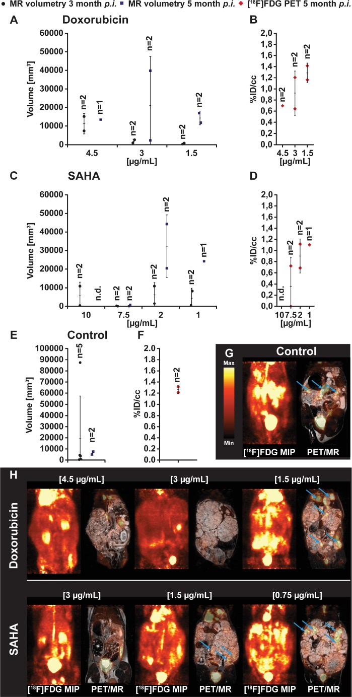
Cytostatic drugs used in cancer therapy were evaluated for their capacity to inhibit metacestode growth and proliferation. Metacestode tissues were exposed to docetaxel, doxorubicin, navelbine, paclitaxel, and vorinostat for 1 week, then incubated in drug-free culture, and thereafter metacestodes were injected into the peritoneum of . Magnetic resonance imaging (MRI) and simultaneous positron emission tomography (PET) were applied to monitor growth of drug-exposed in . At 3 month p.i., docetaxel (at 10 μM, 5 μM and 2 μM) inhibited growth and proliferation of , and at 5 months p.i., only in the 2 μM docetaxel exposure group 0.3 cm of parasite tissue was found. With paclitaxel and navelbine the growth of metacestodes was suppressed until 3 months p.i., thereafter, parasite tissues enlarged up to 3 cm in both groups. tissues of more than 10 g developed in injected with metacestodes which were previously exposed to doxorubicin, navelbine, paclitaxel or vorinostat. In infected with metacestodes previously exposed to docetaxel, the grown parasite tissues weighted 0.2 g. cultured metacestodes exposed to docetaxel did not produce vesicles until 7 weeks post drug exposure, while metacestodes exposed to doxorubicin, navelbine and vorinostat proliferated continuously. In summary, docetaxel, and less efficaciously paclitaxel, inhibited and parasite growth and proliferation, and these observations suggest further experimental studies with selected drug combinations which may translate into new treatment options against alveolar echinococcosis.
对癌症治疗中使用的细胞毒性药物抑制囊尾蚴生长和增殖的能力进行了评估。将囊尾蚴组织暴露于多西他赛、阿霉素、长春瑞滨、紫杉醇和伏立诺他中1周,然后在无药物的培养基中孵育,之后将囊尾蚴注射到……的腹膜中。应用磁共振成像(MRI)和同步正电子发射断层扫描(PET)监测在……中药物处理后的囊尾蚴生长情况。感染后3个月,多西他赛(10μM、5μM和2μM)抑制了……的生长和增殖,感染后5个月,仅在2μM多西他赛处理组发现了0.3cm的寄生虫组织。使用紫杉醇和长春瑞滨时,囊尾蚴的生长在感染后3个月内受到抑制,此后,两组的寄生虫组织均增大至3cm。在注射了先前暴露于阿霉素、长春瑞滨、紫杉醇或伏立诺他的囊尾蚴的……中,出现了超过10g的组织。在感染了先前暴露于多西他赛的囊尾蚴的……中,生长的寄生虫组织重0.2g。暴露于多西他赛的培养囊尾蚴在药物暴露后7周才产生囊泡,而暴露于阿霉素、长春瑞滨和伏立诺他的囊尾蚴则持续增殖。总之,多西他赛以及效果稍差的紫杉醇抑制了……和寄生虫的生长和增殖,这些观察结果表明,对选定的药物组合进行进一步的实验研究可能会转化为针对肺泡型棘球蚴病的新治疗方案。