Division of Pulmonology, Allergy and Critical Care Medicine, Department of Internal Medicine, Korea University College of Medicine, Seoul, Korea.
Department of Physiology, Korea University College of Medicine, Seoul, Korea.
PLoS One. 2018 Mar 8;13(3):e0193117. doi: 10.1371/journal.pone.0193117. eCollection 2018.
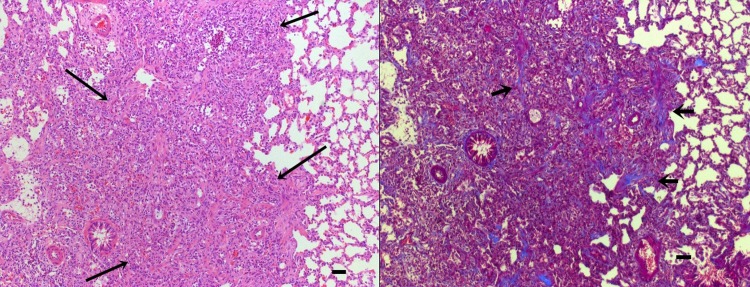
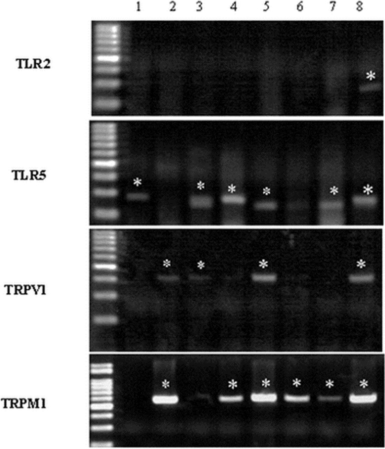
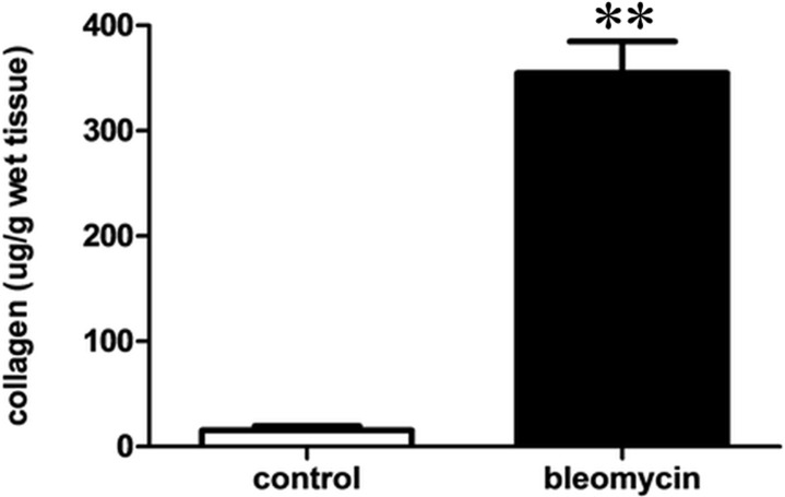
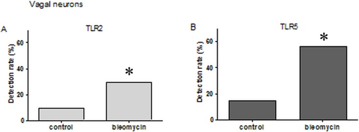
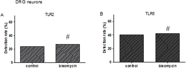
Airway sensory nerves are known to express several receptors and channels that are activated by exogenous and endogenous mediators that cause coughing. Toll-like receptor (TLR) s are expressed in nociceptive neurons and play an important role in neuroinflammation. However, there have been very few studies of TLR expression in lung-derived sensory neurons or their relevance to respiratory symptoms such as cough. We used the bleomycin-induced pulmonary fibrosis model to investigate the change in TLR expression in pulmonary neurons and the association of TLRs with transient receptor potential (TRP) channels in pulmonary neurons. After 2 weeks of bleomycin or saline administration, pulmonary fibrosis changes were confirmed using tissue staining and the SIRCOL collagen assay. TLRs (TLR 1-9) and TRP channel expression was analyzed using single cell reverse transcription polymerase chain reaction (RT-PCR) in isolated sensory neurons from the nodose/jugular ganglion and the dorsal root ganglion (DRG). Pulmonary sensory neurons expressed TLR2 and TLR5. In the bleomycin-induced pulmonary fibrosis model, TLR2 expression was detected in 29.5% (18/61) and 26.9% (21/78) of pulmonary nodose/jugular neurons and DRG neurons, respectively. TLR5 was also detected in 55.7% (34/61) and 42.3% (33/78) of pulmonary nodose/jugular neurons and DRG neurons, respectively, in the bleomycin-induced pulmonary fibrosis model. TLR5 was expressed in 63.4% of TRPV1 positive cells and 43.4% of TRPM8 positive cells. In conclusion, TLR2 and TLR5 expression is enhanced, especially in vagal neurons, in the bleomycin-induced fibrosis model group when compared to the saline treated control group. Co-expression of TLR5 and TRP channels in pulmonary sensory neurons was also observed. This work sheds new light on the role of TLRs in the control and manifestation of clinical symptoms, such as cough. To understand the role of TLRs in pulmonary sensory nerves, further study will be required.
气道感觉神经被认为表达几种受体和通道,这些受体和通道被外源性和内源性介质激活,导致咳嗽。Toll 样受体 (TLR) 在伤害性神经元中表达,并在神经炎症中发挥重要作用。然而,关于肺源性感觉神经元中 TLR 的表达及其与咳嗽等呼吸症状的相关性的研究很少。我们使用博来霉素诱导的肺纤维化模型来研究肺神经元中 TLR 表达的变化,以及 TLR 与肺神经元中瞬时受体电位 (TRP) 通道的相关性。在博来霉素或生理盐水给药 2 周后,通过组织染色和 SIRCOL 胶原测定法确认肺纤维化变化。使用单细胞逆转录聚合酶链反应 (RT-PCR) 分析分离自结状神经节和背根神经节 (DRG) 的感觉神经元中的 TLR (TLR 1-9) 和 TRP 通道表达。肺感觉神经元表达 TLR2 和 TLR5。在博来霉素诱导的肺纤维化模型中,TLR2 表达分别在肺结状/颈神经节和 DRG 神经元中的 29.5%(18/61)和 26.9%(21/78)中检测到。TLR5 也分别在博来霉素诱导的肺纤维化模型中的肺结状/颈神经节和 DRG 神经元中的 55.7%(34/61)和 42.3%(33/78)中检测到。TLR5 在 63.4%的 TRPV1 阳性细胞和 43.4%的 TRPM8 阳性细胞中表达。总之,与生理盐水处理的对照组相比,TLR2 和 TLR5 的表达在博来霉素诱导的纤维化模型组中增强,尤其是在迷走神经神经元中。还观察到 TLR5 和 TRP 通道在肺感觉神经元中的共表达。这项工作为 TLR 在控制和表现临床症状(如咳嗽)方面的作用提供了新的认识。为了了解 TLR 在肺感觉神经中的作用,需要进一步的研究。