Department of Thoracic Cardiovascular Surgery, Affiliated Hospital of Xuzhou Medical University, Xuzhou, Jiangsu 221006, P.R. China.
Research Facility Center for Morphology, Xuzhou Medical University, Xuzhou, Jiangsu 221004, P.R. China.
Int J Mol Med. 2018 Jun;41(6):3243-3252. doi: 10.3892/ijmm.2018.3552. Epub 2018 Mar 9.
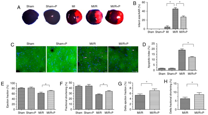
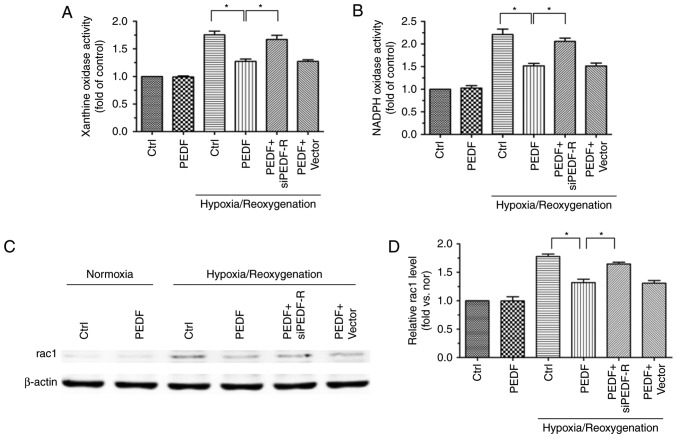
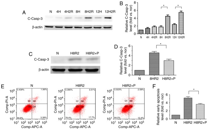
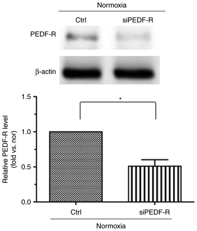
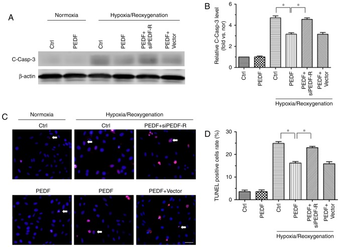
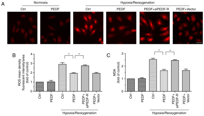
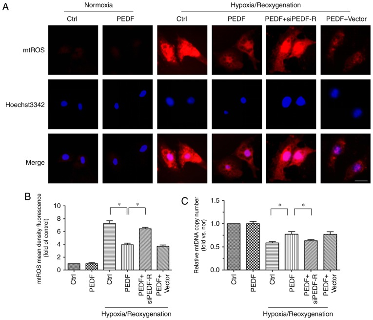
The prevention and management of myocardial ischemia/reperfusion (MI/R) injury is an essential part of coronary heart disease surgery and is becoming a major clinical problem in the treatment of ischemic heart disease. Previous studies by our group have demonstrated that pigment epithelium‑derived factor (PEDF) improves cardiac function in rats with acute myocardial infarction and reduces hypoxia‑induced cell injury. However, the protective function and mechanisms underlying the effect of PEDF in MI/R injury remain to be fully understood. In the present study, the positive effect of PEDF in MI/R injury was confirmed by construction of the adult Sprague‑Dawley rat MI/R model. PEDF reduced myocardial infarct size and downregulated cardiomyocyte apoptosis in the I/R myocardium in this model. In addition, PEDF improved cardiac function and increased cardiac functional reserve in rats subjected to MI/R Injury. To further study the protective effect of PEDF and the underlying mechanisms in MI/R injury, a H9c2 cardiomyocyte hypoxia/reoxygenation (H/R) model was constructed. PEDF was confirmed to decrease H/R‑induced apoptosis in H9c2 cells, and this anti‑apoptotic function was abolished by pigment epithelium‑derived factor‑receptor (PEDF R) small interfering (si)RNA. Furthermore, administration of PEDF decreased the levels of reactive oxygen species (ROS) and malondialdehyde (MDA) in H/R H9c2 cells. Compared with the H/R group, PEDF decreased mitochondrial ROS, increased the mitochondrial DNA copy number, reduced xanthine oxidase and NADPH oxidase activity, as well as RAC family small GTPase 1 protein expression. However, these effects of PEDF were markedly attenuated by PEDF‑R siRNA. To the best of our knowledge, the present study is the first to identify the protective effect of PEDF in MI/R injury, and confirm that the antioxidative effect PEDF occurred via inhibition of ROS generation via PEDF‑R under MI/R conditions.
心肌缺血/再灌注(MI/R)损伤的预防和管理是冠心病手术的重要组成部分,也是缺血性心脏病治疗中的一个主要临床问题。本研究组先前的研究表明,色素上皮衍生因子(PEDF)可改善急性心肌梗死大鼠的心脏功能,并减轻低氧诱导的细胞损伤。然而,PEDF 在 MI/R 损伤中的保护作用及其机制仍有待充分阐明。在本研究中,通过构建成年 Sprague-Dawley 大鼠 MI/R 模型证实了 PEDF 在 MI/R 损伤中的积极作用。PEDF 减少了该模型中 I/R 心肌中的心肌梗死面积并下调了心肌细胞凋亡。此外,PEDF 改善了 MI/R 损伤大鼠的心脏功能并增加了心脏功能储备。为了进一步研究 PEDF 在 MI/R 损伤中的保护作用及其潜在机制,构建了 H9c2 心肌细胞缺氧/复氧(H/R)模型。证实 PEDF 可减少 H9c2 细胞中的 H/R 诱导的细胞凋亡,而这种抗凋亡作用被色素上皮衍生因子受体(PEDF R)小干扰(si)RNA 所消除。此外,给予 PEDF 可降低 H/R H9c2 细胞中的活性氧(ROS)和丙二醛(MDA)水平。与 H/R 组相比,PEDF 降低了线粒体 ROS,增加了线粒体 DNA 拷贝数,减少了黄嘌呤氧化酶和 NADPH 氧化酶活性,以及 RAC 家族小 GTP 酶 1 蛋白表达。然而,PEDF-R siRNA 明显减弱了 PEDF 的这些作用。据我们所知,本研究首次鉴定了 PEDF 在 MI/R 损伤中的保护作用,并证实了在 MI/R 条件下,PEDF 通过抑制 PEDF-R 产生的 ROS 来发挥抗氧化作用。