Laboratório de Toxinologia, Instituto Oswaldo Cruz, FIOCRUZ, Rio de Janeiro RJ 21040-900, Brazil.
Instituto Nacional de Ciência e Tecnologia em Toxinas (INCTTox), CNPq, Brasília DF 71605-170, Brazil.
Toxins (Basel). 2018 Mar 13;10(3):121. doi: 10.3390/toxins10030121.
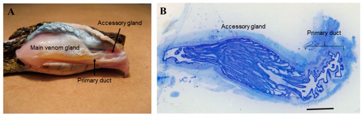
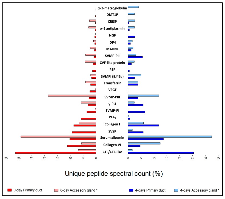
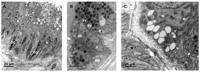
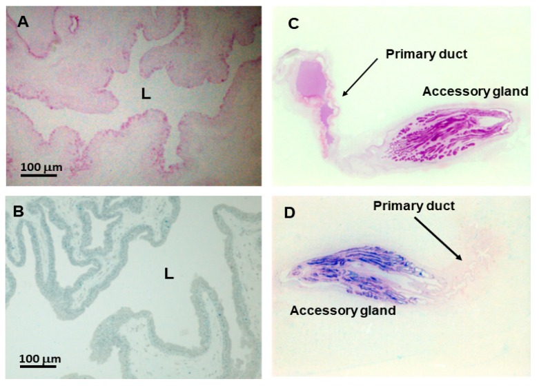
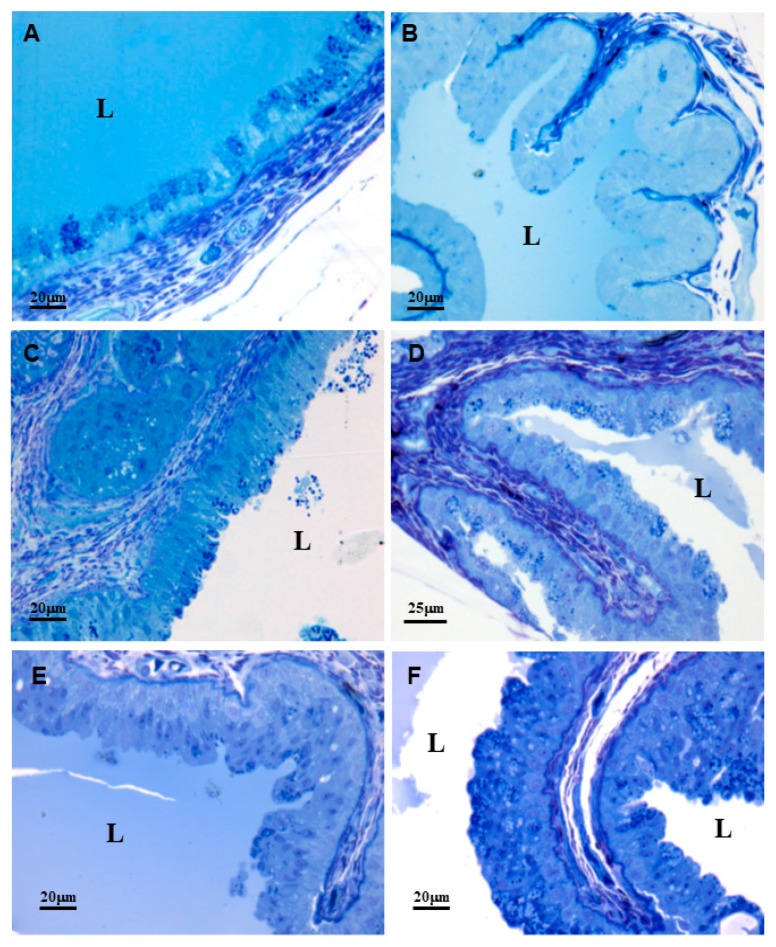
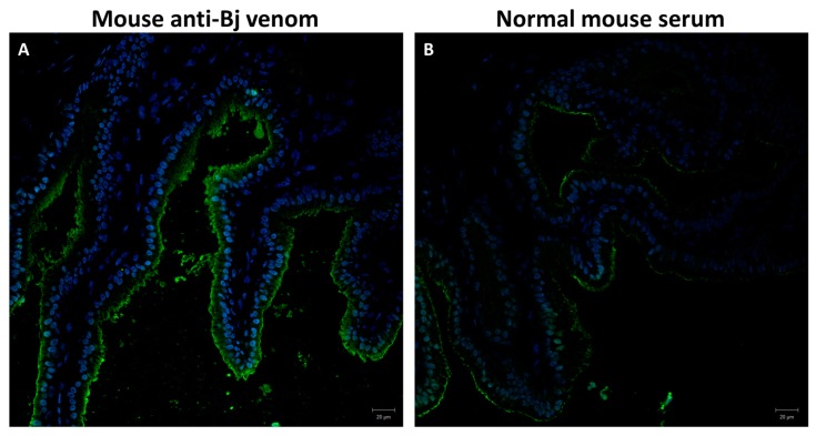
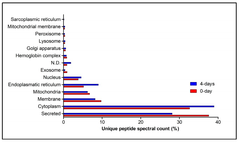
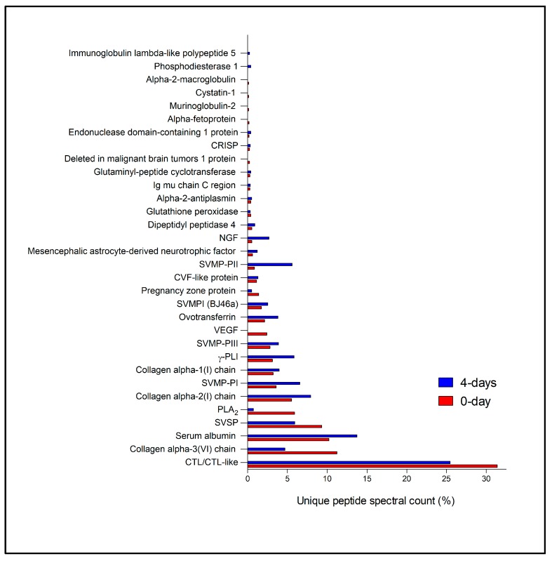
Despite numerous studies concerning morphology and venom production and secretion in the main venom gland (and some data on the accessory gland) of the venom glandular apparatus of Viperidae snakes, the primary duct has been overlooked. We characterized the primary duct of the snake by morphological analysis, immunohistochemistry and proteomics. The duct has a pseudostratified epithelium with secretory columnar cells with vesicles of various electrondensities, as well as mitochondria-rich, dark, basal, and horizontal cells. Morphological analysis, at different periods after venom extraction, showed that the primary duct has a long cycle of synthesis and secretion, as do the main venom and accessory glands; however, the duct has a mixed mode venom storage, both in the lumen and in secretory vesicles. Mouse anti- venom serum strongly stained the primary duct's epithelium. Subsequent proteomic analysis revealed the synthesis of venom toxins-mainly C-type lectin/C-type lectin-like proteins. We propose that the primary duct's toxin synthesis products complement the final venom bolus. Finally, we hypothesize that the primary duct and the accessory gland (components of the venom glandular apparatus) are part of the evolutionary path from a salivary gland towards the main venom gland.
尽管已经有许多关于蝰蛇科蛇类毒液腺的主要毒液腺(以及一些关于附属腺的资料)的形态学和毒液产生与分泌的研究,但初级导管一直被忽视。我们通过形态分析、免疫组织化学和蛋白质组学来描述蛇的初级导管。该导管具有假复层上皮,分泌柱状细胞,含有各种电子密度的囊泡,以及富含线粒体的深棕色基底细胞和平行细胞。形态分析显示,在毒液提取后的不同时期,初级导管具有与主毒液腺和附属腺相同的长合成和分泌周期;然而,导管具有混合模式的毒液储存方式,既在管腔中也在分泌小泡中。小鼠抗毒液血清强烈染色初级导管的上皮。随后的蛋白质组学分析显示了毒液毒素的合成-主要是 C 型凝集素/C 型凝集素样蛋白。我们提出,初级导管的毒素合成产物补充了最终的毒液团块。最后,我们假设初级导管和附属腺(毒液腺器官的组成部分)是从唾液腺向主毒液腺进化过程的一部分。