Karolinska Institute, Department of Neurobiology, Care Sciences and Society, Division of Neurogeriatrics, Center for Alzheimer Research, Huddinge, Sweden.
Division of Neurodegeneration, Huddinge, Sweden.
J Cell Mol Med. 2018 Jun;22(6):3016-3024. doi: 10.1111/jcmm.13534. Epub 2018 Mar 13.
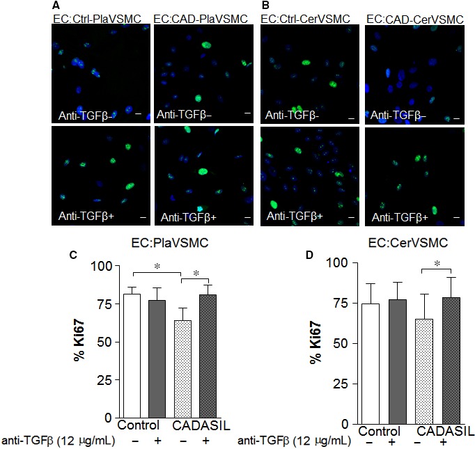
Cerebral autosomal-dominant arteriopathy with subcortical infarcts and leukoencephalopathy (CADASIL) is a familial fatal progressive degenerative disorder. One of the pathological hallmarks of CADASIL is a dramatic reduction of vascular smooth muscle cells (VSMCs) in cerebral arteries. Using VSMCs from the vasculature of the human umbilical cord, placenta and cerebrum of CADASIL patients, we found that CADASIL VSMCs had a lower proliferation rate compared to control VSMCs. Exposure of control VSMCs and endothelial cells (ECs) to media derived from CADASIL VSMCs lowered the proliferation rate of all cells examined. By quantitative RT-PCR analysis, we observed increased Transforming growth factor-β (TGFβ) gene expression in CADASIL VSMCs. Adding TGFβ-neutralizing antibody restored the proliferation rate of CADASIL VSMCs. We assessed proliferation differences in the presence or absence of TGFβ-neutralizing antibody in ECs co-cultured with VSMCs. ECs co-cultured with CADASIL VSMCs exhibited a lower proliferation rate than those co-cultured with control VSMCs, and neutralization of TGFβ normalized the proliferation rate of ECs co-cultured with CADASIL VSMCs. We suggest that increased TGFβ expression in CADASIL VSMCs is involved in the reduced VSMC proliferation in CADASIL and may play a role in situ in altered proliferation of neighbouring cells in the vasculature.
脑常染色体显性遗传性动脉病伴皮质下梗死和白质脑病(CADASIL)是一种家族性致命性进行性退行性疾病。CADASIL 的病理标志之一是大脑动脉中的血管平滑肌细胞(VSMCs)明显减少。使用来自 CADASIL 患者血管、胎盘和大脑的血管中的 VSMCs,我们发现与对照 VSMCs 相比,CADASIL VSMCs 的增殖率较低。将对照 VSMCs 和内皮细胞(ECs)暴露于源自 CADASIL VSMCs 的培养基中,降低了所有被检测细胞的增殖率。通过定量 RT-PCR 分析,我们观察到 CADASIL VSMCs 中转化生长因子-β(TGFβ)基因表达增加。添加 TGFβ 中和抗体恢复了 CADASIL VSMCs 的增殖率。我们评估了在存在或不存在 TGFβ 中和抗体的情况下,ECs 与 VSMCs 共培养时的增殖差异。与对照 VSMCs 共培养的 ECs 的增殖率低于与 CADASIL VSMCs 共培养的 ECs,中和 TGFβ 使与 CADASIL VSMCs 共培养的 ECs 的增殖率正常化。我们认为,CADASIL VSMCs 中 TGFβ 表达增加参与了 CADASIL 中 VSMC 增殖减少,并可能在血管中邻近细胞增殖改变的原位发挥作用。