• 文献检索
  • 文档翻译
  • 深度研究
  • 学术资讯
  • Suppr Zotero 插件Zotero 插件
  • 邀请有礼
  • 套餐&价格
  • 历史记录
应用&插件
Suppr Zotero 插件Zotero 插件浏览器插件Mac 客户端Windows 客户端微信小程序
定价
高级版会员购买积分包购买API积分包
服务
文献检索文档翻译深度研究API 文档MCP 服务
关于我们
关于 Suppr公司介绍联系我们用户协议隐私条款
关注我们

Suppr 超能文献

核心技术专利:CN118964589B侵权必究
粤ICP备2023148730 号-1Suppr @ 2026

文献检索

告别复杂PubMed语法,用中文像聊天一样搜索,搜遍4000万医学文献。AI智能推荐,让科研检索更轻松。

立即免费搜索

文件翻译

保留排版,准确专业,支持PDF/Word/PPT等文件格式,支持 12+语言互译。

免费翻译文档

深度研究

AI帮你快速写综述,25分钟生成高质量综述,智能提取关键信息,辅助科研写作。

立即免费体验

胆汁淤积性肝损伤中的细胞间相互作用

Cellular crosstalk during cholestatic liver injury.

作者信息

Thomson Joanne, Hargrove Laura, Kennedy Lindsey, Demieville Jennifer, Francis Heather

机构信息

Research, Central Texas Veterans Healthcare System, TX, USA.

Medicine, Texas A&M Health Science Center, Temple, TX, USA.

出版信息

Liver Res. 2017 Jun;1(1):26-33. doi: 10.1016/j.livres.2017.05.002. Epub 2017 May 10.

DOI:10.1016/j.livres.2017.05.002
PMID:29552372
原文链接:https://pmc.ncbi.nlm.nih.gov/articles/PMC5854144/
Abstract

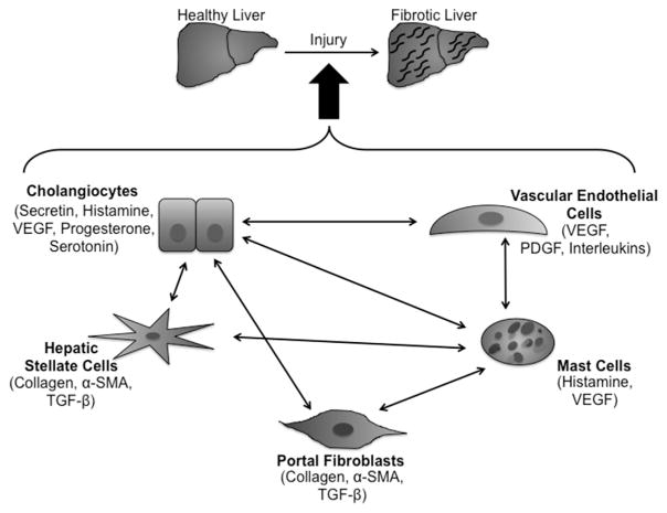
The functions of the liver are very diverse. From detoxifying blood to storing glucose in the form of glycogen and producing bile to facilitate fat digestion, the liver is a very active and important organ. The liver is comprised of many varied cell types whose functions are equally diverse. Cholangiocytes line the biliary tree and aid in transporting and adjusting the composition of bile as it travels to the gallbladder. Hepatic stellate cells and portal fibroblasts are located in different areas within the liver architecture, but both contribute to the development of fibrosis upon activation after liver injury. Vascular cells, including those that constitute the peribiliary vascular plexus, are involved in functions other than blood delivery to and from the liver, such as supporting the growth of the biliary tree during development. Mast cells are normally found in healthy livers but in very low numbers. However, after injury, mast cell numbers greatly increase as they infiltrate and release factors that exacerbate the fibrotic response. While not an all-inclusive list, these cells have individual roles within the liver, but they are also able to communicate with each other by cellular crosstalk. In this review, we examine some of these pathways that can lead to an increase in the homeostatic dysfunction seen in liver injury.

摘要

肝脏的功能多种多样。从对血液进行解毒,到以糖原的形式储存葡萄糖,以及产生胆汁以促进脂肪消化,肝脏是一个非常活跃且重要的器官。肝脏由许多不同类型的细胞组成,其功能同样多样。胆管细胞排列在胆管树中,在胆汁输送至胆囊的过程中,协助运输并调节胆汁的成分。肝星状细胞和门静脉成纤维细胞位于肝脏结构内的不同区域,但在肝损伤后激活时,二者都会促使肝纤维化的发展。血管细胞,包括构成胆小管周围血管丛的细胞,除了参与肝脏的血液进出输送外,还具有其他功能,比如在发育过程中支持胆管树的生长。肥大细胞通常存在于健康肝脏中,但数量极少。然而在损伤后,肥大细胞数量会大幅增加,它们会浸润并释放加剧纤维化反应的因子。虽然这并非全部细胞类型,但这些细胞在肝脏中各自发挥着作用,而且它们还能够通过细胞间相互作用进行交流。在本综述中,我们研究了其中一些可能导致肝损伤中稳态功能障碍加剧的途径。

https://cdn.ncbi.nlm.nih.gov/pmc/blobs/207c/5854144/8d27bea065cc/nihms946997f2.jpg
https://cdn.ncbi.nlm.nih.gov/pmc/blobs/207c/5854144/f0a38fe0a9d7/nihms946997f1.jpg
https://cdn.ncbi.nlm.nih.gov/pmc/blobs/207c/5854144/8d27bea065cc/nihms946997f2.jpg
https://cdn.ncbi.nlm.nih.gov/pmc/blobs/207c/5854144/f0a38fe0a9d7/nihms946997f1.jpg
https://cdn.ncbi.nlm.nih.gov/pmc/blobs/207c/5854144/8d27bea065cc/nihms946997f2.jpg

相似文献

1
Cellular crosstalk during cholestatic liver injury.胆汁淤积性肝损伤中的细胞间相互作用
Liver Res. 2017 Jun;1(1):26-33. doi: 10.1016/j.livres.2017.05.002. Epub 2017 May 10.
2
Cooperation of liver cells in health and disease.健康与疾病状态下肝细胞的协作。
Adv Anat Embryol Cell Biol. 2001;161:III-XIII, 1-151. doi: 10.1007/978-3-642-56553-3.
3
Hedgehog Signaling Demarcates a Niche of Fibrogenic Peribiliary Mesenchymal Cells.刺猬信号标志着纤维生成性胆周间充质细胞的龛位。
Gastroenterology. 2020 Aug;159(2):624-638.e9. doi: 10.1053/j.gastro.2020.03.075. Epub 2020 Apr 11.
4
Peribiliary myofibroblasts in biliary type liver fibrosis.胆汁型肝纤维化中的胆管周围肌成纤维细胞
Front Biosci. 2002 Feb 1;7:d496-503. doi: 10.2741/A790.
5
Activated hepatic stellate cells and portal fibroblasts contribute to cholestatic liver fibrosis in MDR2 knockout mice.活化的肝星状细胞和门脉成纤维细胞有助于 MDR2 敲除小鼠的胆汁淤积性肝纤维化。
J Hepatol. 2019 Sep;71(3):573-585. doi: 10.1016/j.jhep.2019.04.012. Epub 2019 May 7.
6
Danhongqing formula alleviates cholestatic liver fibrosis by downregulating long non-coding RNA H19 derived from cholangiocytes and inhibiting hepatic stellate cell activation.丹红清方通过下调胆管细胞来源的长链非编码RNA H19和抑制肝星状细胞活化来减轻胆汁淤积性肝纤维化。
J Integr Med. 2024 Mar;22(2):188-198. doi: 10.1016/j.joim.2024.03.006. Epub 2024 Mar 9.
7
The characteristics of activated portal fibroblasts/myofibroblasts in liver fibrosis.肝纤维化中活化的肝门成纤维细胞/肌成纤维细胞的特征
Differentiation. 2016 Sep;92(3):84-92. doi: 10.1016/j.diff.2016.07.001. Epub 2016 Aug 31.
8
Fibrotic Events in the Progression of Cholestatic Liver Disease.胆汁淤积性肝病进展中的纤维化事件。
Cells. 2021 May 5;10(5):1107. doi: 10.3390/cells10051107.
9
Distribution of intrahepatic mast cells in various hepatobiliary disorders. An immunohistochemical study.各种肝胆疾病中肝内肥大细胞的分布。一项免疫组织化学研究。
Virchows Arch. 1998 Nov;433(5):471-9. doi: 10.1007/s004280050276.
10
Bile duct ligation-induced biliary hyperplasia, hepatic injury, and fibrosis are reduced in mast cell-deficient Kit mice.在肥大细胞缺陷的Kit小鼠中,胆管结扎诱导的胆管增生、肝损伤和纤维化有所减轻。
Hepatology. 2017 Jun;65(6):1991-2004. doi: 10.1002/hep.29079. Epub 2017 Apr 28.

引用本文的文献

1
Gut-liver axis: Recent concepts in pathophysiology in alcohol-associated liver disease.肠-肝轴:酒精性肝病病理生理学的最新概念
Hepatology. 2024 Dec 1;80(6):1342-1371. doi: 10.1097/HEP.0000000000000924. Epub 2024 May 1.
2
Immune-mediated cholangiopathies in children: the need to better understand the pathophysiology for finding the future possible treatment targets.儿童免疫介导性胆管病:为寻找未来可能的治疗靶点,需要更好地了解其病理生理学。
Front Immunol. 2023 Oct 20;14:1206025. doi: 10.3389/fimmu.2023.1206025. eCollection 2023.
3
Indole-3-Carboxaldehyde Restores Gut Mucosal Integrity and Protects from Liver Fibrosis in Murine Sclerosing Cholangitis.

本文引用的文献

1
Vascular endothelial growth factor promotes the activation of hepatic stellate cells in chronic schistosomiasis.血管内皮生长因子促进慢性血吸虫病中肝星状细胞的活化。
Immunol Cell Biol. 2017 Apr;95(4):399-407. doi: 10.1038/icb.2016.109. Epub 2016 Nov 3.
2
Endothelial cells: source, barrier, and target of defensive mediators.内皮细胞:防御介质的来源、屏障和靶标。
Immunol Rev. 2016 Nov;274(1):307-329. doi: 10.1111/imr.12479.
3
Knockout of microRNA-21 reduces biliary hyperplasia and liver fibrosis in cholestatic bile duct ligated mice.
吲哚-3-甲醛可修复肠道黏膜完整性并防止小鼠硬化性胆管炎肝纤维化。
Cells. 2021 Jun 29;10(7):1622. doi: 10.3390/cells10071622.
4
New insights on the role of vascular endothelial growth factor in biliary pathophysiology.血管内皮生长因子在胆汁病理生理学中作用的新见解。
JHEP Rep. 2021 Feb 4;3(3):100251. doi: 10.1016/j.jhepr.2021.100251. eCollection 2021 Jun.
5
Stimulation of hepatocarcinogenesis by activated cholangiocytes via Il17a/f1 pathway in kras transgenic zebrafish model.激活的胆管细胞通过 Kras 转基因斑马鱼模型中的 Il17a/f1 通路刺激肝癌发生。
Sci Rep. 2021 Jan 14;11(1):1372. doi: 10.1038/s41598-020-80621-6.
6
Mast Cells in Liver Fibrogenesis.肝纤维化中的肥大细胞。
Cells. 2019 Nov 13;8(11):1429. doi: 10.3390/cells8111429.
7
Protein tyrosine phosphatase 1b deficiency protects against hepatic fibrosis by modulating nadph oxidases.蛋白酪氨酸磷酸酶 1b 缺乏通过调节 NADPH 氧化酶来防止肝纤维化。
Redox Biol. 2019 Sep;26:101263. doi: 10.1016/j.redox.2019.101263. Epub 2019 Jun 29.
8
Amelioration of Ductular Reaction by Stem Cell Derived Extracellular Vesicles in MDR2 Knockout Mice via Lethal-7 microRNA.通过致死因子-7 微 RNA,干细胞来源的细胞外囊泡改善 MDR2 敲除小鼠胆管反应。
Hepatology. 2019 Jun;69(6):2562-2578. doi: 10.1002/hep.30542. Epub 2019 Apr 12.
9
Liver-Derived Exosomes and Their Implications in Liver Pathobiology.肝脏来源的外泌体及其在肝脏病理生物学中的意义。
Int J Mol Sci. 2018 Nov 22;19(12):3715. doi: 10.3390/ijms19123715.
10
Hepatic Cytochrome P450 Activity and Nitric Oxide Production During Multiple Ovalbumin Challenges.多次卵清蛋白激发过程中肝脏细胞色素P450活性及一氧化氮生成情况
Eur J Drug Metab Pharmacokinet. 2019 Jun;44(3):379-387. doi: 10.1007/s13318-018-0527-1.
敲除微小RNA-21可减轻胆管结扎诱导的胆汁淤积性小鼠的胆管增生和肝纤维化。
Lab Invest. 2016 Dec;96(12):1256-1267. doi: 10.1038/labinvest.2016.112. Epub 2016 Oct 24.
4
Collagen-binding vascular endothelial growth factor attenuates CCl4-induced liver fibrosis in mice.胶原结合血管内皮生长因子减轻小鼠四氯化碳诱导的肝纤维化。
Mol Med Rep. 2016 Nov;14(5):4680-4686. doi: 10.3892/mmr.2016.5826. Epub 2016 Oct 12.
5
Forkhead box A2 regulates biliary heterogeneity and senescence during cholestatic liver injury in mice‡.叉头框A2调节小鼠胆汁淤积性肝损伤期间的胆管异质性和衰老‡
Hepatology. 2017 Feb;65(2):544-559. doi: 10.1002/hep.28831. Epub 2016 Nov 5.
6
Biology of Cholangiocytes: From Bench to Bedside.胆管细胞生物学:从 bench 到 bedside 。 (注:bench 常表示基础研究阶段,bedside 常表示临床应用阶段,这里表示从基础研究到临床应用)
Gut Liver. 2016 Sep 15;10(5):687-98. doi: 10.5009/gnl16033.
7
Isolation and characterization of hepatic mast cells from cholestatic rats.胆汁淤积性大鼠肝肥大细胞的分离与鉴定
Lab Invest. 2016 Nov;96(11):1198-1210. doi: 10.1038/labinvest.2016.89. Epub 2016 Aug 22.
8
Inhibition of mast cell-secreted histamine decreases biliary proliferation and fibrosis in primary sclerosing cholangitis Mdr2(-/-) mice.抑制肥大细胞分泌的组胺可减少原发性硬化性胆管炎Mdr2(-/-)小鼠的胆管增殖和纤维化。
Hepatology. 2016 Oct;64(4):1202-1216. doi: 10.1002/hep.28704. Epub 2016 Jul 30.
9
Mast Cell-Targeted Strategies in Cancer Therapy.癌症治疗中的肥大细胞靶向策略。
Transfus Med Hemother. 2016 Mar;43(2):109-13. doi: 10.1159/000444942. Epub 2016 Mar 9.
10
Effect of Mast Cell Chymase on Activation, Proliferation and Transdifferentiation of Hepatic Stellate Cells.肥大细胞糜蛋白酶对肝星状细胞激活、增殖和转分化的影响
Hepatogastroenterology. 2015 Jun;62(140):1007-10.