Philadelphia College of Osteopathic Medicine, Philadelphia, PA, USA.
John A. Moran Eye Center, University of Utah, Salt Lake City, UT, USA.
Exp Eye Res. 2018 Jun;171:101-105. doi: 10.1016/j.exer.2018.03.016. Epub 2018 Mar 17.
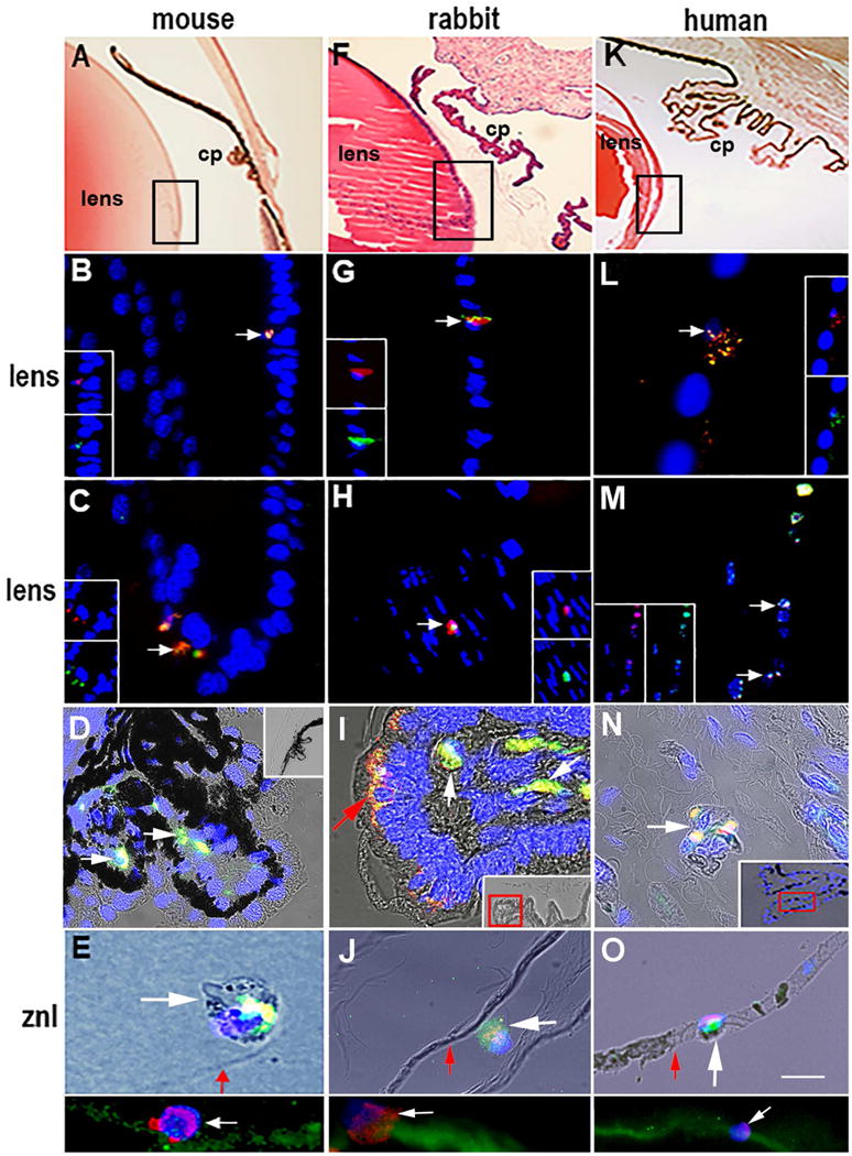
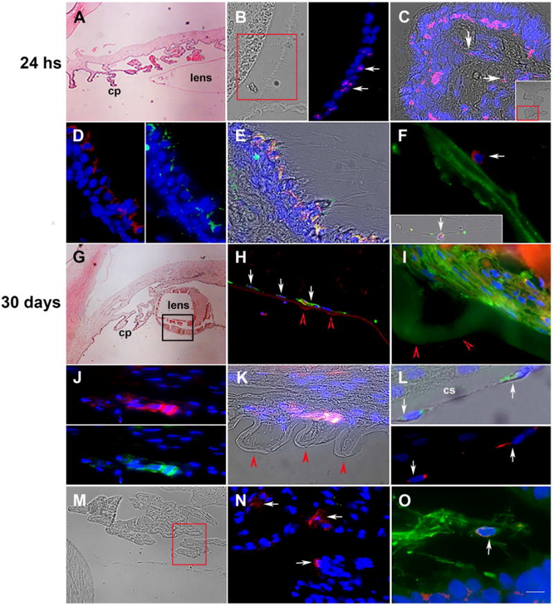
Myo/Nog cells, named for their expression of MyoD and noggin, enter the eye during early stages of embryonic development. Their release of noggin is critical for normal morphogenesis of the lens and retina. Myo/Nog cells are also present in adult eyes. Single nucleated skeletal muscle cells designated as myofibroblasts arise from Myo/Nog cells in cultures of lens tissue. In this report we document the presence of Myo/Nog cells in the lens, ciliary body and on the zonule of Zinn in mice, rabbits and humans. Myo/Nog cells were rare in all three structures. Their prevalence increased in the lens and ciliary body of rabbits 24 h following cataract surgery. Rabbits developed posterior capsule opacification (PCO) within one month of surgery. The number of Myo/Nog cells continued to be elevated in the lens and ciliary body. Myo/Nog cells containing alpha smooth muscle actin and striated muscle myosin were present on the posterior capsule and overlaid deformations in the capsule. Myo/Nog cells also were present on the zonule fibers and external surface of the posterior capsule. These findings suggest that Myo/Nog contribute to PCO and may use the zonule fibers to migrate between the ciliary processes and lens.
肌源性/诺格细胞因其表达 MyoD 和诺金而得名,在胚胎发育的早期进入眼睛。它们释放诺金对于晶状体和视网膜的正常形态发生至关重要。肌源性/诺格细胞也存在于成年眼睛中。在晶状体组织的培养物中,来自肌源性/诺格细胞的单核骨骼肌细胞被指定为肌成纤维细胞。在本报告中,我们记录了肌源性/诺格细胞在小鼠、兔子和人类晶状体、睫状体和 Zinn 带中的存在。在这三种结构中,肌源性/诺格细胞都很少见。在白内障手术后 24 小时,兔子的晶状体和睫状体中,肌源性/诺格细胞的患病率增加。兔子在手术后一个月内发展为后囊混浊(PCO)。晶状体和睫状体中的肌源性/诺格细胞数量继续升高。在后囊和覆盖在囊上的变形处存在含有α平滑肌肌动蛋白和横纹肌肌球蛋白的肌源性/诺格细胞。肌源性/诺格细胞也存在于悬韧带纤维和后囊的外表面。这些发现表明肌源性/诺格细胞有助于 PCO 的发生,并且可能利用悬韧带纤维在睫状体和晶状体之间迁移。