Suppr超能文献

女性骨盆扩散加权成像的陷阱

Pitfalls of diffusion-weighted imaging of the female pelvis.

作者信息

Duarte Ana Luisa, Dias João Lopes, Cunha Teresa Margarida

机构信息

Department of Radiology, Hospital do Espírito Santo E.P.E., Évora, Portugal.

Department of Radiology, Hospital de São José, Centro Hospitalar de Lisboa Central, Lisboa, Portugal.

出版信息

Radiol Bras. 2018 Jan-Feb;51(1):37-44. doi: 10.1590/0100-3984.2016.0208.

Abstract

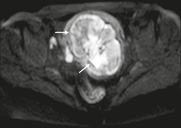
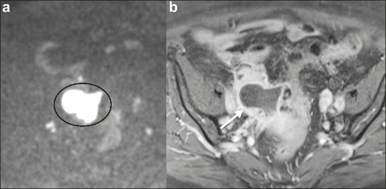
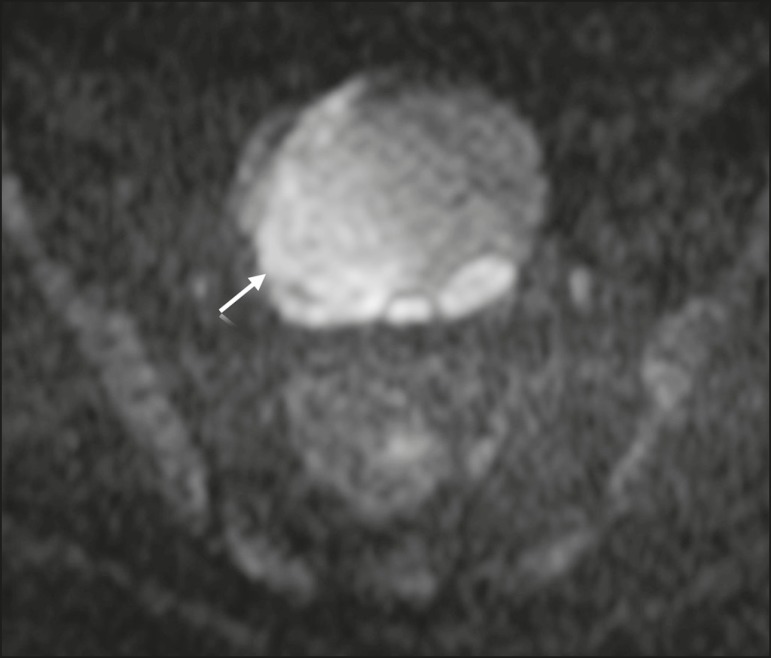
Diffusion-weighted imaging (DWI) is widely used in protocols for magnetic resonance imaging (MRI) of the female pelvis. It provides functional and structural information about biological tissues, without the use of ionizing radiation or intravenous administration of contrast medium. High signal intensity on DWI with simultaneous low signal intensity on apparent diffusion coefficient maps is usually associated with malignancy. However, that pattern can also be seen in many benign lesions, a fact that should be recognized by radiologists. Correlating DWI findings with those of conventional (T1- and T2-weighted) MRI sequences and those of contrast-enhanced MRI sequences is mandatory in order to avoid potential pitfalls. The aim of this review article is the description of the most relevant physiological and benign pathological conditions of the female pelvis that can show restricted diffusion on DWI.

摘要

扩散加权成像(DWI)广泛应用于女性盆腔磁共振成像(MRI)方案中。它无需使用电离辐射或静脉注射造影剂就能提供有关生物组织的功能和结构信息。DWI上的高信号强度与同时出现的表观扩散系数图上的低信号强度通常与恶性肿瘤有关。然而,这种表现模式在许多良性病变中也可见,放射科医生应认识到这一事实。为避免潜在的陷阱,必须将DWI的结果与传统(T1加权和T2加权)MRI序列以及对比增强MRI序列的结果进行关联。这篇综述文章的目的是描述女性盆腔中在DWI上可显示扩散受限的最相关生理和良性病理状况。

https://cdn.ncbi.nlm.nih.gov/pmc/blobs/8c2b/5846323/4136e9e44351/rb-51-01-0037-g01.jpg

相似文献

1
Pitfalls of diffusion-weighted imaging of the female pelvis.
Radiol Bras. 2018 Jan-Feb;51(1):37-44. doi: 10.1590/0100-3984.2016.0208.
4
Non-neoplastic diseases of the fallopian tube: MR imaging with emphasis on diffusion-weighted imaging.
Insights Imaging. 2016 Jun;7(3):311-27. doi: 10.1007/s13244-016-0484-7. Epub 2016 Mar 18.
10
The role of diffusion weighted magnetic resonance imaging in oncologic settings.
Quant Imaging Med Surg. 2013 Oct;3(5):269-78. doi: 10.3978/j.issn.2223-4292.2013.10.07.

引用本文的文献

1
An Update on the Role of MRI in Treatment Stratification of Patients with Cervical Cancer.
Cancers (Basel). 2023 Oct 23;15(20):5105. doi: 10.3390/cancers15205105.
2
[Adnexal Masses: Clinical Application of Multiparametric MR Imaging & O-RADS MRI].
Taehan Yongsang Uihakhoe Chi. 2021 Sep;82(5):1066-1082. doi: 10.3348/jksr.2021.0111. Epub 2021 Sep 15.
3
Utility of the Diffusion Weighted Sequence in Gynecological Imaging: Review Article.
Cancers (Basel). 2022 Sep 15;14(18):4468. doi: 10.3390/cancers14184468.
4
Acute pelvic pain: A pictorial review with magnetic resonance imaging.
J Clin Imaging Sci. 2022 Aug 17;12:48. doi: 10.25259/JCIS_70_2022. eCollection 2022.
5
The role of diffusion-weighted imaging in the evaluation of adnexal lesions.
Pol J Radiol. 2022 Aug 20;87:e469-e477. doi: 10.5114/pjr.2022.119064. eCollection 2022.
6
Diagnostic Value and Clinical Application of Diffusion-Weighted Magnetic Resonance Imaging for Female Pelvic Lesions.
Contrast Media Mol Imaging. 2022 Jun 20;2022:5868453. doi: 10.1155/2022/5868453. eCollection 2022.
7
Spontaneous round ligament hematoma as an unusual cause of pelvic pain in a young female patient: MRI demonstration.
Radiol Case Rep. 2022 Mar 25;17(5):1765-1769. doi: 10.1016/j.radcr.2022.02.079. eCollection 2022 May.
8
10
Transvaginal ultrasound in deep endometriosis: pictorial essay.
Radiol Bras. 2019 Sep-Oct;52(5):337-341. doi: 10.1590/0100-3984.2018.0019.

本文引用的文献

2
Non-oncologic applications of diffusion-weighted imaging (DWI) in the genitourinary system.
Abdom Imaging. 2015 Aug;40(6):1645-54. doi: 10.1007/s00261-015-0471-3.
3
The shading sign: is it exclusive of endometriomas?
Abdom Imaging. 2015 Oct;40(7):2566-72. doi: 10.1007/s00261-015-0465-1.
5
The utility of diffusion-weighted magnetic resonance imaging in differentiation of endometriomas from hemorrhagic ovarian cysts.
Clin Imaging. 2015 Sep-Oct;39(5):830-3. doi: 10.1016/j.clinimag.2015.05.003. Epub 2015 May 7.
7
Differentiation of ovarian endometriomas from hemorrhagic cysts at MR imaging: utility of the T2 dark spot sign.
Radiology. 2014 Apr;271(1):126-32. doi: 10.1148/radiol.13131394. Epub 2013 Nov 13.
8
Pearls and pitfalls in MRI of gynecologic malignancy with diffusion-weighted technique.
AJR Am J Roentgenol. 2013 Feb;200(2):261-76. doi: 10.2214/AJR.12.9713.
10
MR imaging of endometriosis: ten imaging pearls.
Radiographics. 2012 Oct;32(6):1675-91. doi: 10.1148/rg.326125518.

文献AI研究员

20分钟写一篇综述,助力文献阅读效率提升50倍。

立即体验

用中文搜PubMed

大模型驱动的PubMed中文搜索引擎

马上搜索

文档翻译

学术文献翻译模型,支持多种主流文档格式。

立即体验