Department of Psychology, Leiden University, Leiden, The Netherlands.
Leiden Institute for Brain and Cognition, Leiden, The Netherlands.
J Child Psychol Psychiatry. 2018 Sep;59(9):994-1002. doi: 10.1111/jcpp.12895. Epub 2018 Mar 25.
Adolescence is a transition period characterized by heightened emotional reactivity, which for some sets the stage for emerging depressive symptoms. Prior studies suggest that adolescent depression is associated with deviant cortical and subcortical brain structure. Longitudinal studies are, however, currently scarce, but critical to detect which adolescents are at risk for developing depressive symptoms.
In this longitudinal study, a community sample of 205 participants underwent magnetic resonance imaging (MRI) in three biennial waves (522 scans) spanning 5 years across ages 8-25 years. Depressive symptomatology was assessed using self-report at the third time point. Mixed models were used to examine the relations between structural brain development, specifically regional change in cortical thickness, surface area and subcortical volumes (hippocampus and amygdala), and depressive symptoms.
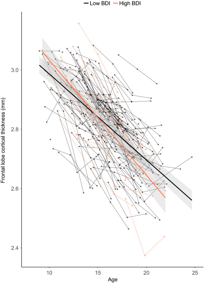
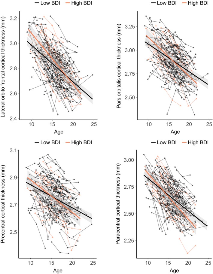
Accelerated frontal lobe cortical thinning was observed in adolescents who developed depressive symptoms at the third time point. This effect remained after controlling for parent-reported affective problems at the first time point. Moreover, the effect was driven by specific lateral orbitofrontal and precentral regions. In addition, differential developmental trajectories of parietal cortical thickness and surface area in several regions were found for participants reporting higher depressive symptomatology, but these results did not survive correction for multiple comparisons. Volumes or developmental volume changes in hippocampus or amygdala were not related to depressive symptoms.
This study showed that emerging depression is associated with cortical thinning in frontal regions within individuals. These findings move beyond detecting cross-sectional correlations and set the stage for early detection, which may inform future intervention.
青春期是一个情绪反应强烈的过渡时期,对于一些人来说,这为出现抑郁症状奠定了基础。先前的研究表明,青少年抑郁与皮质和皮质下脑结构的异常有关。然而,纵向研究目前还很少,但对于检测哪些青少年有出现抑郁症状的风险至关重要。
在这项纵向研究中,一个社区样本的 205 名参与者在三个两年一次的波次(522 次扫描)中接受了磁共振成像(MRI),跨越了 5 年的年龄范围,从 8 岁到 25 岁。在第三次时间点使用自我报告评估抑郁症状。使用混合模型来检查结构脑发育(特别是皮质厚度、表面积和皮质下体积(海马体和杏仁核)的区域变化)与抑郁症状之间的关系。
在第三次时间点出现抑郁症状的青少年中,观察到额前皮质变薄加速。在控制第一次时间点父母报告的情感问题后,这一效应仍然存在。此外,这种效应是由特定的外侧眶额和中央前区驱动的。此外,对于报告更高抑郁症状的参与者,发现了几个区域的顶叶皮质厚度和表面积的差异发展轨迹,但这些结果在进行多次比较校正后并不成立。海马体或杏仁核的体积或发育体积变化与抑郁症状无关。
这项研究表明,新出现的抑郁与个体额前区域的皮质变薄有关。这些发现超越了检测横断面相关性,并为早期检测奠定了基础,这可能为未来的干预提供信息。