Biophysical Analytics, German Rheumatism Research Center, 10117 Berlin, Germany.
Neuropathology, Charité-Universitätsmedizin, 10117 Berlin, Germany.
Int J Mol Sci. 2018 Mar 29;19(4):1018. doi: 10.3390/ijms19041018.
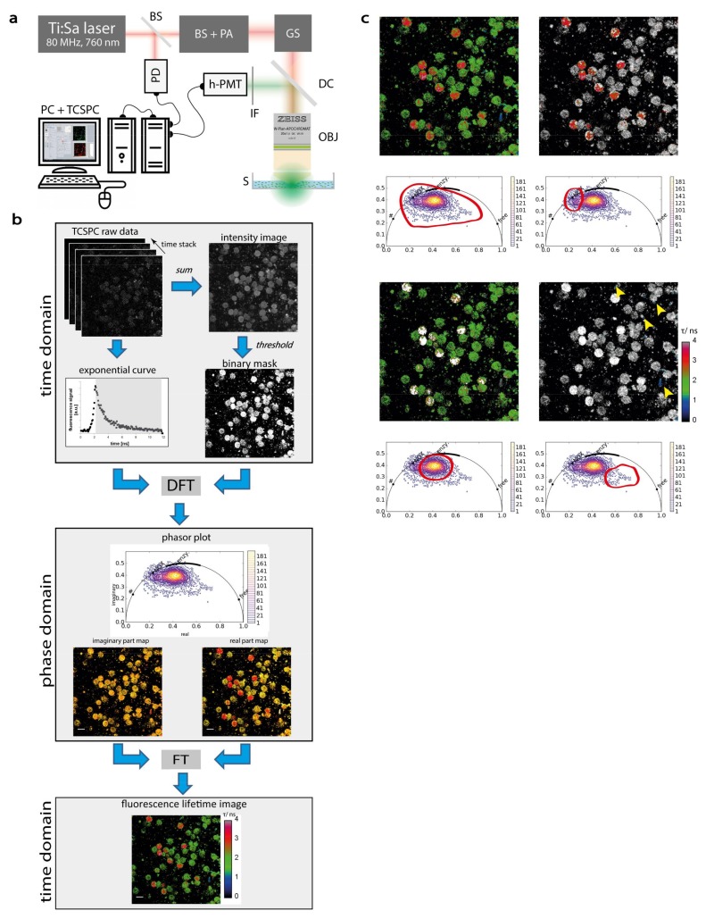
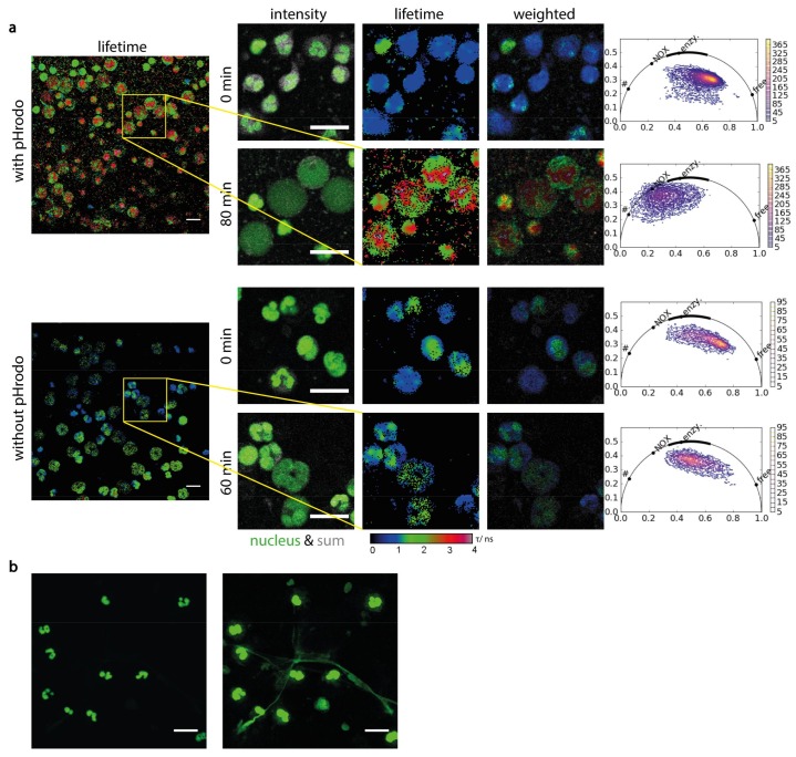
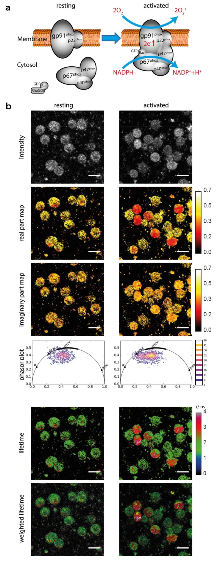
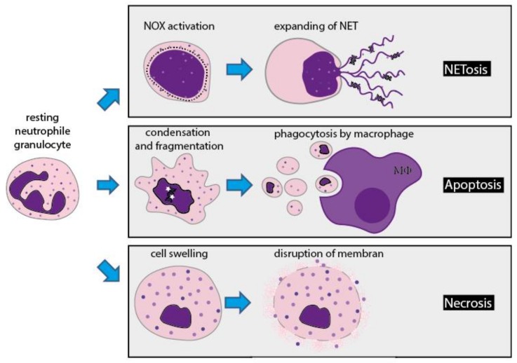
Time-correlated single-photon counting combined with multi-photon laser scanning microscopy has proven to be a versatile tool to perform fluorescence lifetime imaging in biological samples and, thus, shed light on cellular functions, both in vitro and in vivo. Here, by means of phasor-analyzed endogenous NAD(P)H (nicotinamide adenine dinucleotide (phosphate)) fluorescence lifetime imaging, we visualize the shift in the cellular metabolism of healthy human neutrophil granulocytes during phagocytosis of pHrodo™ beads. We correlate this with the process of NETosis, i.e., trapping of pathogens by DNA networks. Hence, we are able to directly show the dynamics of NADPH oxidase activation and its requirement in triggering NETosis in contrast to other pathways of cell death and to decipher the dedicated spatio-temporal sequence between NADPH oxidase activation, nuclear membrane disintegration and DNA network formation. The endogenous FLIM approach presented here uniquely meets the increasing need in the field of immunology to monitor cellular metabolism as a basic mechanism of cellular and tissue functions.
时间相关单光子计数与多光子激光扫描显微镜相结合,已被证明是一种在生物样本中进行荧光寿命成像的多功能工具,从而揭示了细胞功能,无论是在体外还是体内。在这里,通过相分析内源性 NAD(P)H(烟酰胺腺嘌呤二核苷酸(磷酸))荧光寿命成像,我们可以观察到健康人嗜中性粒细胞吞噬 pHrodo™珠时细胞代谢的变化。我们将其与 NETosis 过程(即通过 DNA 网络捕获病原体)相关联。因此,我们能够直接显示 NADPH 氧化酶激活的动力学及其在触发 NETosis 方面与细胞死亡的其他途径的要求,并破译 NADPH 氧化酶激活、核膜崩解和 DNA 网络形成之间的特定时空序列。这里提出的内源性 FLIM 方法独特地满足了免疫学领域作为细胞和组织功能基本机制监测细胞代谢的日益增长的需求。