• 文献检索
  • 文档翻译
  • 深度研究
  • 学术资讯
  • Suppr Zotero 插件Zotero 插件
  • 邀请有礼
  • 套餐&价格
  • 历史记录
应用&插件
Suppr Zotero 插件Zotero 插件浏览器插件Mac 客户端Windows 客户端微信小程序
定价
高级版会员购买积分包购买API积分包
服务
文献检索文档翻译深度研究API 文档MCP 服务
关于我们
关于 Suppr公司介绍联系我们用户协议隐私条款
关注我们

Suppr 超能文献

核心技术专利:CN118964589B侵权必究
粤ICP备2023148730 号-1Suppr @ 2026

文献检索

告别复杂PubMed语法,用中文像聊天一样搜索,搜遍4000万医学文献。AI智能推荐,让科研检索更轻松。

立即免费搜索

文件翻译

保留排版,准确专业,支持PDF/Word/PPT等文件格式,支持 12+语言互译。

免费翻译文档

深度研究

AI帮你快速写综述,25分钟生成高质量综述,智能提取关键信息,辅助科研写作。

立即免费体验

基于 NAD(P)H 的双光子荧光寿命成像显微镜技术对单个细胞外囊泡进行无标记特征分析。

Label-free characterization of single extracellular vesicles using two-photon fluorescence lifetime imaging microscopy of NAD(P)H.

机构信息

Beckman Institute for Advanced Science and Technology, University of Illinois at Urbana-Champaign, Urbana, IL, 61801, USA.

Department of Bioengineering, University of Illinois at Urbana-Champaign, Urbana, IL, 61801, USA.

出版信息

Sci Rep. 2021 Feb 8;11(1):3308. doi: 10.1038/s41598-020-80813-0.

DOI:10.1038/s41598-020-80813-0
PMID:33558561
原文链接:https://pmc.ncbi.nlm.nih.gov/articles/PMC7870923/
Abstract

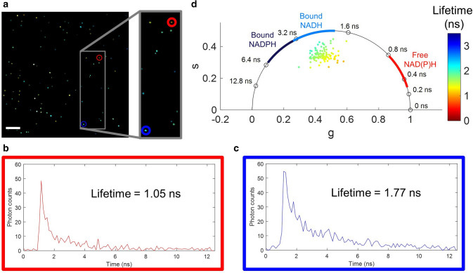
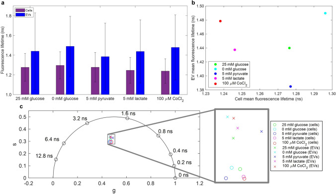
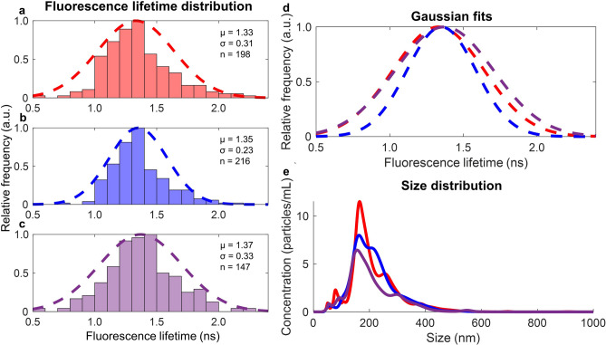
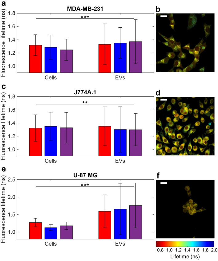
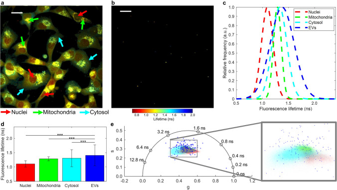
The heterogeneous nature of extracellular vesicles (EVs) creates the need for single EV characterization techniques. However, many common biochemical and functional EV analysis techniques lack single EV resolution. Two-photon fluorescence lifetime imaging microscopy (FLIM) is widely used to functionally characterize the reduced form of nicotinamide adenine dinucleotide and nicotinamide adenine dinucleotide phosphate (NAD(P)H) in cells and tissues. Here, we demonstrate that FLIM can also be used to image and characterize NAD(P)H in single isolated EVs. EVs were isolated using standard differential ultracentrifugation techniques from multiple cell lines and imaged using a custom two-photon FLIM system. The presented data show that the NAD(P)H fluorescence lifetimes in isolated cell-derived EVs follow a wide Gaussian distribution, indicating the presence of a range of different protein-bound and free NAD(P)H species. EV NAD(P)H fluorescence lifetime distribution has a larger standard deviation than that of cells and a significantly different fluorescence lifetime distribution than the nuclei, mitochondria, and cytosol of cells. Additionally, changes in the metabolic conditions of cells were reflected in changes in the mean fluorescence lifetime of NAD(P)H in the produced EVs. These data suggest that FLIM of NAD(P)H could be a valuable tool for EV research.

摘要

细胞外囊泡 (EVs) 的异质性要求采用单一 EV 特征分析技术。然而,许多常见的生化和功能 EV 分析技术缺乏对单一 EV 的分辨率。双光子荧光寿命成像显微镜 (FLIM) 广泛用于对细胞和组织中还原型烟酰胺腺嘌呤二核苷酸 (NAD[H]) 和烟酰胺腺嘌呤二核苷酸磷酸 (NAD[P]H) 的功能进行特征分析。在这里,我们证明 FLIM 也可用于对单个分离的 EV 中的 NAD(P)H 进行成像和特征分析。使用标准差速超速离心技术从多种细胞系中分离 EV,并使用定制的双光子 FLIM 系统对其进行成像。所呈现的数据表明,分离的细胞衍生 EV 中的 NAD(P)H 荧光寿命遵循宽高斯分布,表明存在一系列不同的蛋白结合和游离 NAD(P)H 物质。EV NAD(P)H 荧光寿命分布的标准偏差大于细胞的标准偏差,并且与细胞的核、线粒体和细胞质的荧光寿命分布显著不同。此外,细胞代谢条件的变化反映在产生的 EV 中 NAD(P)H 的平均荧光寿命的变化上。这些数据表明,NAD(P)H 的 FLIM 可能是 EV 研究的一个有价值的工具。

https://cdn.ncbi.nlm.nih.gov/pmc/blobs/71e2/7870923/56e00444e18e/41598_2020_80813_Fig6_HTML.jpg
https://cdn.ncbi.nlm.nih.gov/pmc/blobs/71e2/7870923/927bd1fd88c7/41598_2020_80813_Fig1_HTML.jpg
https://cdn.ncbi.nlm.nih.gov/pmc/blobs/71e2/7870923/abd52a06ecdc/41598_2020_80813_Fig2_HTML.jpg
https://cdn.ncbi.nlm.nih.gov/pmc/blobs/71e2/7870923/cde408ef2ba7/41598_2020_80813_Fig3_HTML.jpg
https://cdn.ncbi.nlm.nih.gov/pmc/blobs/71e2/7870923/da047f7ecab4/41598_2020_80813_Fig4_HTML.jpg
https://cdn.ncbi.nlm.nih.gov/pmc/blobs/71e2/7870923/efff657f2835/41598_2020_80813_Fig5_HTML.jpg
https://cdn.ncbi.nlm.nih.gov/pmc/blobs/71e2/7870923/56e00444e18e/41598_2020_80813_Fig6_HTML.jpg

相似文献

1
Label-free characterization of single extracellular vesicles using two-photon fluorescence lifetime imaging microscopy of NAD(P)H.基于 NAD(P)H 的双光子荧光寿命成像显微镜技术对单个细胞外囊泡进行无标记特征分析。
Sci Rep. 2021 Feb 8;11(1):3308. doi: 10.1038/s41598-020-80813-0.
2
Multiphoton FLIM imaging of NAD(P)H and FAD with one excitation wavelength.利用单一激发波长的多光子 FLIM 成像技术对 NAD(P)H 和 FAD 进行成像。
J Biomed Opt. 2020 Jan;25(1):1-16. doi: 10.1117/1.JBO.25.1.014510.
3
Simultaneous assessment of NAD(P)H and flavins with multispectral fluorescence lifetime imaging microscopy at a single excitation wavelength of 750 nm.在 750nm 的单一激发波长下,利用多光谱荧光寿命成像显微镜同时评估 NAD(P)H 和黄素。
J Biomed Opt. 2024 Oct;29(10):106501. doi: 10.1117/1.JBO.29.10.106501. Epub 2024 Sep 30.
4
Two-photon FLIM of NAD(P)H and FAD in mesenchymal stem cells undergoing either osteogenic or chondrogenic differentiation.对正在经历成骨或软骨分化的间充质干细胞中烟酰胺腺嘌呤二核苷酸(磷酸)(NAD(P)H)和黄素腺嘌呤二核苷酸(FAD)进行双光子荧光寿命成像。
Stem Cell Res Ther. 2017 Jan 28;8(1):15. doi: 10.1186/s13287-017-0484-7.
5
Development and characterization of phasor-based analysis for FLIM to evaluate the metabolic and epigenetic impact of HER2 inhibition on squamous cell carcinoma cultures.基于相量分析的 FLIM 开发与特征描述,用于评估 HER2 抑制对鳞状细胞癌细胞培养物的代谢和表观遗传影响。
J Biomed Opt. 2021 Oct;26(10). doi: 10.1117/1.JBO.26.10.106501.
6
Two-photon fluorescence lifetime imaging monitors metabolic changes during wound healing of corneal epithelial cells in vitro.双光子荧光寿命成像监测体外角膜上皮细胞愈合过程中的代谢变化。
Graefes Arch Clin Exp Ophthalmol. 2012 Sep;250(9):1293-302. doi: 10.1007/s00417-012-2051-3. Epub 2012 May 6.
7
Label-free fluorescence lifetime imaging for the assessment of cell viability in living tumor fragments.无标记荧光寿命成像用于评估活肿瘤组织片段中的细胞活力。
J Biomed Opt. 2024 Jun;29(Suppl 2):S22709. doi: 10.1117/1.JBO.29.S2.S22709. Epub 2024 Jun 14.
8
NAD(P)H fluorescence lifetime measurements in fixed biological tissues.固定生物组织中的 NAD(P)H 荧光寿命测量。
Methods Appl Fluoresc. 2019 Oct 10;7(4):044005. doi: 10.1088/2050-6120/ab47e5.
9
Single-cell redox states analyzed by fluorescence lifetime metrics and tryptophan FRET interaction with NAD(P)H.利用荧光寿命指标分析单细胞氧化还原状态及色氨酸与 NAD(P)H 的 FRET 相互作用。
Cytometry A. 2019 Jan;95(1):110-121. doi: 10.1002/cyto.a.23711. Epub 2019 Jan 2.
10
Two-photon fluorescence lifetime imaging of intrinsic NADH in three-dimensional tumor models.三维肿瘤模型中内源性 NADH 的双光子荧光寿命成像。
Cytometry A. 2019 Jan;95(1):80-92. doi: 10.1002/cyto.a.23632. Epub 2018 Oct 21.

引用本文的文献

1
Extracellular Vesicles for Clinical Diagnostics: From Bulk Measurements to Single-Vesicle Analysis.用于临床诊断的细胞外囊泡:从整体测量到单囊泡分析
ACS Nano. 2025 Aug 12;19(31):28021-28109. doi: 10.1021/acsnano.5c00706. Epub 2025 Jul 28.
2
Label-free method to monitor metabolism during long-term culture of human pluripotent stem cell derived cardiomyocytes.用于监测人多能干细胞衍生心肌细胞长期培养过程中代谢的无标记方法。
Biophotonics Discov. 2025 Apr;2(2). doi: 10.1117/1.bios.2.2.025001. Epub 2025 Apr 17.
3
Label-free nonlinear optical signatures of extracellular vesicles in liquid and tissue biopsies of human breast cancer.

本文引用的文献

1
K-means clustering of coherent Raman spectra from extracellular vesicles visualized by label-free multiphoton imaging.无标记多光子成像可视化细胞外囊泡的相干拉曼光谱的 K-均值聚类。
Opt Lett. 2020 Jul 1;45(13):3613-3616. doi: 10.1364/OL.395838.
2
Ca mediates extracellular vesicle biogenesis through alternate pathways in malignancy.钙通过恶性肿瘤中的替代途径介导细胞外囊泡生物发生。
J Extracell Vesicles. 2020 Mar 3;9(1):1734326. doi: 10.1080/20013078.2020.1734326. eCollection 2020.
3
Label-free identification and chemical characterisation of single extracellular vesicles and lipoproteins by synchronous Rayleigh and Raman scattering.
人乳腺癌液体和组织活检中外泌体的无标记非线性光学特征。
Sci Rep. 2024 Mar 6;14(1):5528. doi: 10.1038/s41598-024-55781-4.
4
Label-free approaches for extracellular vesicle detection.用于细胞外囊泡检测的无标记方法。
iScience. 2023 Sep 30;26(11):108105. doi: 10.1016/j.isci.2023.108105. eCollection 2023 Nov 17.
5
Formalin fixation and paraffin embedding interfere with the preservation of optical metabolic assessments based on endogenous NAD(P)H and FAD two-photon excited fluorescence.福尔马林固定和石蜡包埋会干扰基于内源性烟酰胺腺嘌呤二核苷酸(磷酸)(NAD(P)H)和黄素腺嘌呤二核苷酸(FAD)双光子激发荧光的光学代谢评估的保存。
Biomed Opt Express. 2023 Sep 15;14(10):5238-5253. doi: 10.1364/BOE.498297. eCollection 2023 Oct 1.
6
In vivo label-free optical signatures of chemotherapy response in human pancreatic ductal adenocarcinoma patient-derived xenografts.人胰腺导管腺癌患者来源异种移植体化疗反应的体内无标记光学特征。
Commun Biol. 2023 Sep 25;6(1):980. doi: 10.1038/s42003-023-05368-y.
7
A Review of Labeling Approaches Used in Small Extracellular Vesicles Tracing and Imaging.小细胞外囊泡示踪与成像中标记方法的研究综述
Int J Nanomedicine. 2023 Aug 11;18:4567-4588. doi: 10.2147/IJN.S416131. eCollection 2023.
8
Isolation and characterization of extracellular vesicles and future directions in diagnosis and therapy.细胞外囊泡的分离与鉴定及其在诊断与治疗中的未来方向。
Wiley Interdiscip Rev Nanomed Nanobiotechnol. 2023 Jan;15(1):e1835. doi: 10.1002/wnan.1835. Epub 2022 Jul 27.
9
A Criterion of Colorectal Cancer Diagnosis Using Exosome Fluorescence-Lifetime Imaging.一种基于外泌体荧光寿命成像的结直肠癌诊断标准
Diagnostics (Basel). 2022 Jul 24;12(8):1792. doi: 10.3390/diagnostics12081792.
10
Changing Perspectives from Oxidative Stress to Redox Signaling-Extracellular Redox Control in Translational Medicine.转化医学中从氧化应激到氧化还原信号传导——细胞外氧化还原控制的视角转变
Antioxidants (Basel). 2022 Jun 16;11(6):1181. doi: 10.3390/antiox11061181.
通过同步瑞利散射和拉曼散射对单个细胞外囊泡和脂蛋白进行无标记识别及化学表征。
J Extracell Vesicles. 2020 Feb 19;9(1):1730134. doi: 10.1080/20013078.2020.1730134. eCollection 2020.
4
Label-free whole-colony imaging and metabolic analysis of metastatic pancreatic cancer by an autoregulating flexible optical system.无标记全菌落成像和代谢分析转移性胰腺癌的自调节柔性光学系统。
Theranostics. 2020 Jan 1;10(4):1849-1860. doi: 10.7150/thno.40869. eCollection 2020.
5
Tracking metabolic dynamics of apoptosis with high-speed two-photon fluorescence lifetime imaging microscopy.利用高速双光子荧光寿命成像显微镜追踪细胞凋亡的代谢动力学。
Biomed Opt Express. 2019 Nov 21;10(12):6408-6421. doi: 10.1364/BOE.10.006408. eCollection 2019 Dec 1.
6
Proteomic analysis of cerebrospinal fluid extracellular vesicles reveals synaptic injury, inflammation, and stress response markers in HIV patients with cognitive impairment.脑脊液细胞外囊泡的蛋白质组学分析揭示了认知障碍的 HIV 患者中的突触损伤、炎症和应激反应标志物。
J Neuroinflammation. 2019 Dec 5;16(1):254. doi: 10.1186/s12974-019-1617-y.
7
Performance assessment of total RNA sequencing of human biofluids and extracellular vesicles.人体生物体液和细胞外囊泡总 RNA 测序的性能评估。
Sci Rep. 2019 Nov 26;9(1):17574. doi: 10.1038/s41598-019-53892-x.
8
Label-free visualization and characterization of extracellular vesicles in breast cancer.无标记可视化和乳腺癌细胞外囊泡的特征分析。
Proc Natl Acad Sci U S A. 2019 Nov 26;116(48):24012-24018. doi: 10.1073/pnas.1909243116. Epub 2019 Nov 15.
9
Simultaneous label-free autofluorescence and multi-harmonic imaging reveals structural and metabolic changes in murine skin.同步无标记自发荧光和多谐波成像揭示小鼠皮肤的结构和代谢变化。
Biomed Opt Express. 2019 Oct 1;10(10):5431-5444. doi: 10.1364/BOE.10.005431.
10
Imaging of Extracellular Vesicles by Atomic Force Microscopy.通过原子力显微镜对细胞外囊泡进行成像
J Vis Exp. 2019 Sep 11(151). doi: 10.3791/59254.