Jiangsu Key Laboratory of Molecular and Functional Imaging, Department of Radiology, Zhongda Hospital, Medical School of Southeast University, Nanjing.
Key Laboratory of Smart Drug Delivery, Ministry of Education & PLA, School of Pharmacy, Fudan University, Shanghai, People's Republic of China.
Int J Nanomedicine. 2018 Mar 22;13:1819-1829. doi: 10.2147/IJN.S152976. eCollection 2018.
Endothelial progenitor cells (EPCs) play an important role in repairing ischemia tissues. However, the survival, migration and therapeutic efficacy of EPCs after transplantation need to be better understood for further cell therapy.
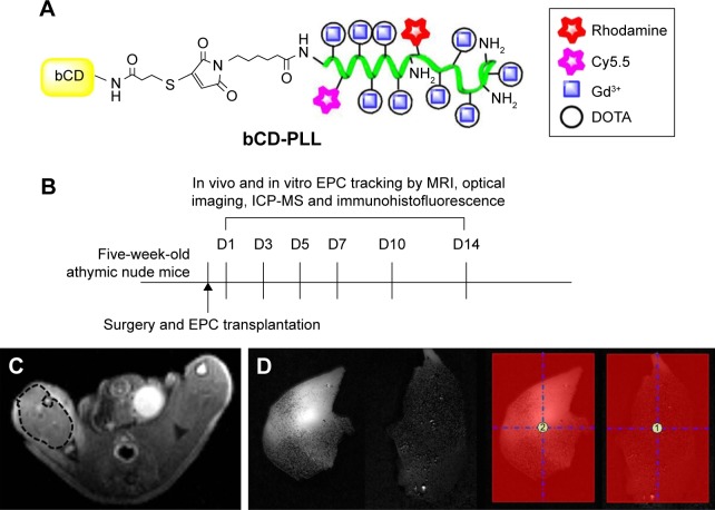
This study investigated the migration effect of EPCs labeled with a multimodal imaging agent in a murine ischemic hindlimb model, using magnetic resonance imaging (MRI) and optical imaging after transplantation.
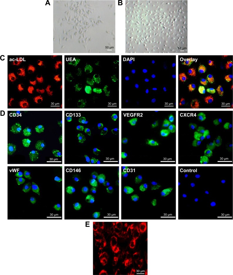
EPCs derived from mouse bone marrow were labeled with a multimodal imaging agent and were administered through intracardiac delivery to mice with ischemic hindlimbs. The injected EPCs and their migration effect were observed via MRI and optical imaging in vivo, and then compared to a reference standard based on histological data. The quantification of gadolinium in tissue samples was done using inductively coupled plasma mass spectrometry (ICP-MS).
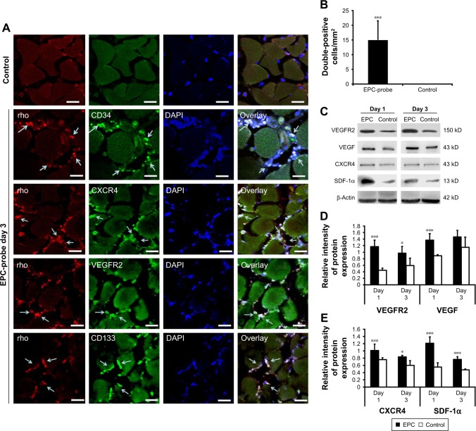
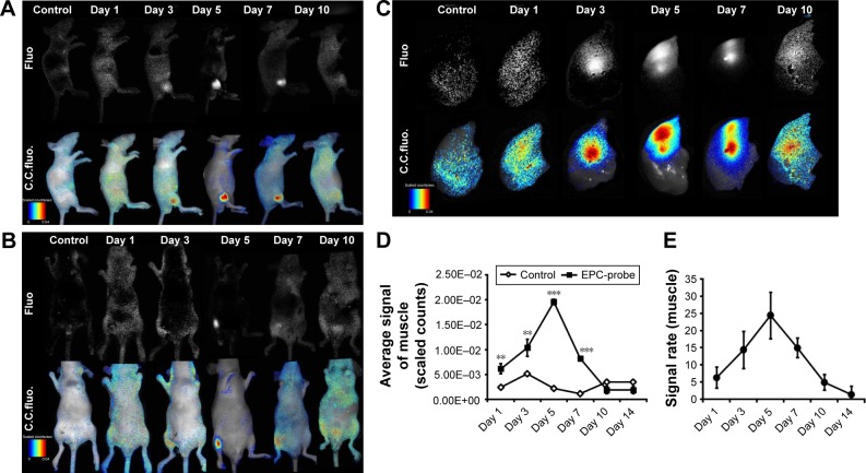
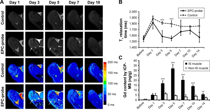
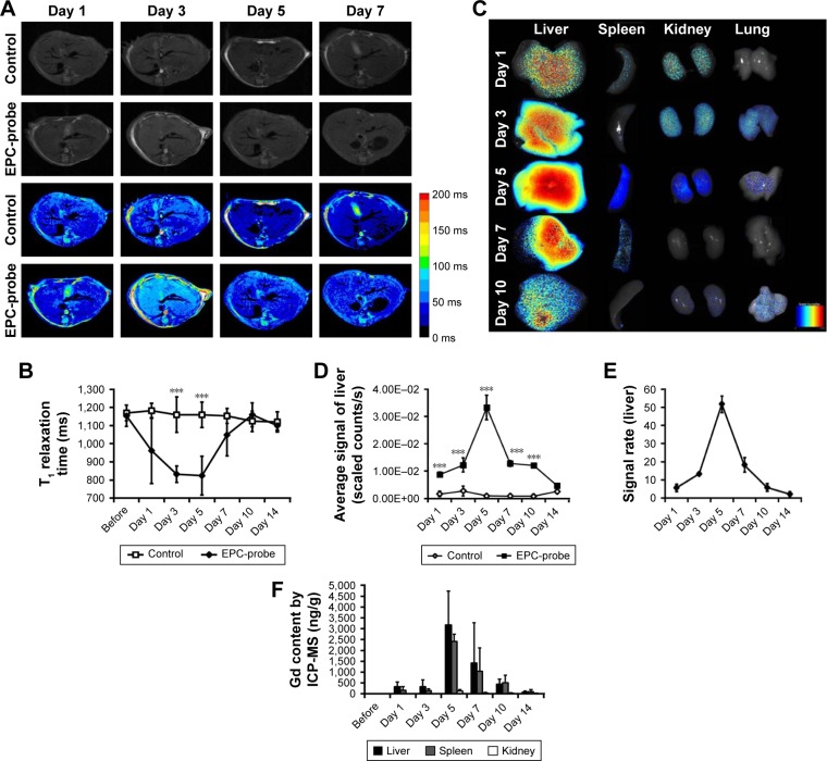
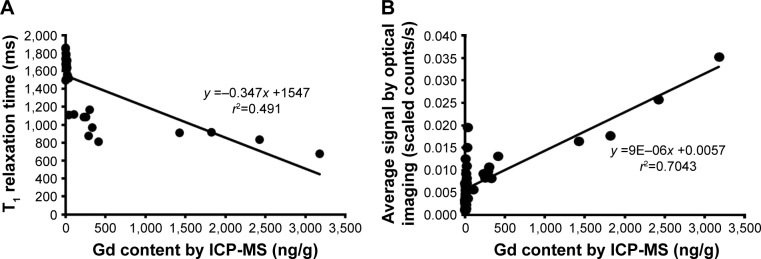
Using in vivo MRI and optical imaging, the labeled EPCs were observed to migrate to ischemic muscle on days 3-5 after injection, while ex vivo, the EPCs were observed in the capillary vessels of the injured tissue. There were significant linear correlations between the Gd contents measured using ICP-MS in samples from the ischemic hindlimbs and livers and T relaxation times calculated using MRI, as well as the average fluorescence signal intensities recorded in optical images (T relaxation time: =0.491; average signal from optical imaging: =0.704, <0.01). EPC treatment upregulated the levels of C-X-C chemokine receptor 4 and vascular endothelial growth factor (VEGF) receptor 2 and enhanced the expression of stromal cell-derived factor-1 and VEGF.
Transplanted EPCs can be monitored with noninvasive MRI and optical imaging in vivo and were found to enhance the paracrine secretion of angiogenic factors.
内皮祖细胞(EPCs)在修复缺血组织中发挥着重要作用。然而,为了进一步的细胞治疗,需要更好地了解移植后 EPCs 的存活、迁移和治疗效果。
本研究通过磁共振成像(MRI)和移植后光学成像,研究了标记有多功能成像剂的 EPC 在小鼠缺血后肢模型中的迁移效应。
从鼠骨髓中分离出 EPCs,并用多功能成像剂进行标记,并通过心内给药将其注射到缺血后肢的小鼠体内。通过体内 MRI 和光学成像观察注射的 EPCs 及其迁移效应,并与基于组织学数据的参考标准进行比较。使用电感耦合等离子体质谱法(ICP-MS)对组织样本中的钆含量进行定量。
通过体内 MRI 和光学成像观察到,标记的 EPCs 在注射后 3-5 天迁移到缺血肌肉,而在体外,EPCs 则存在于受损组织的毛细血管中。ICP-MS 测量的缺血后肢和肝脏样本中的 Gd 含量与 MRI 计算的 T 弛豫时间之间以及光学图像中记录的平均荧光信号强度之间存在显著的线性相关性(T 弛豫时间:=0.491;光学图像中的平均信号:=0.704,<0.01)。EPC 治疗上调了 C-X-C 趋化因子受体 4 和血管内皮生长因子受体 2 的水平,并增强了基质细胞衍生因子-1 和血管内皮生长因子的表达。
移植的 EPCs 可以通过非侵入性的体内 MRI 和光学成像进行监测,并发现它们可以增强旁分泌分泌的血管生成因子。