Weigelin Bettina, Bakker Gert-Jan, Friedl Peter
Department of Cell Biology; Radboud University Nijmegen Medical Centre; Nijmegen, The Netherlands.
David H. Koch Center for Applied Research of Genitourinary Cancers; Department of Genitourinary Medical Oncology; The University of Texas MD Anderson Cancer Center; Houston, TX USA.
Intravital. 2012 Jul 1;1(1):32-43. doi: 10.4161/intv.21223. eCollection 2012.
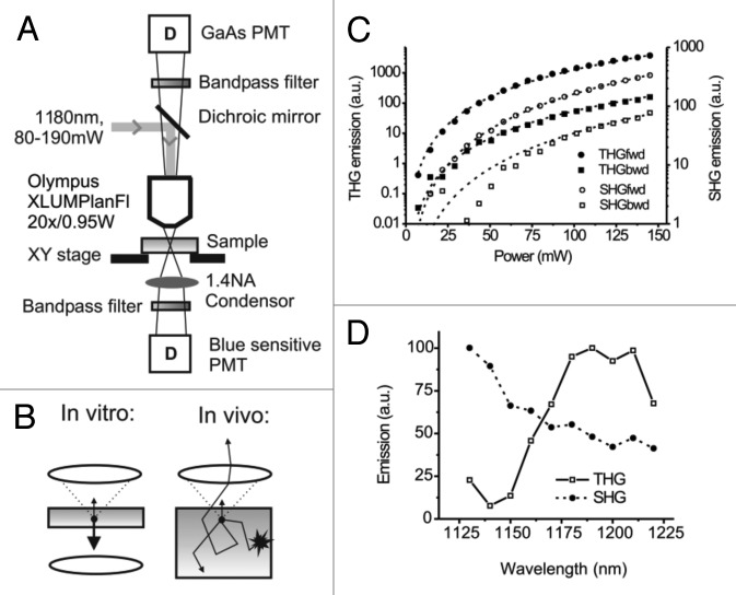
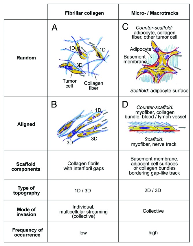
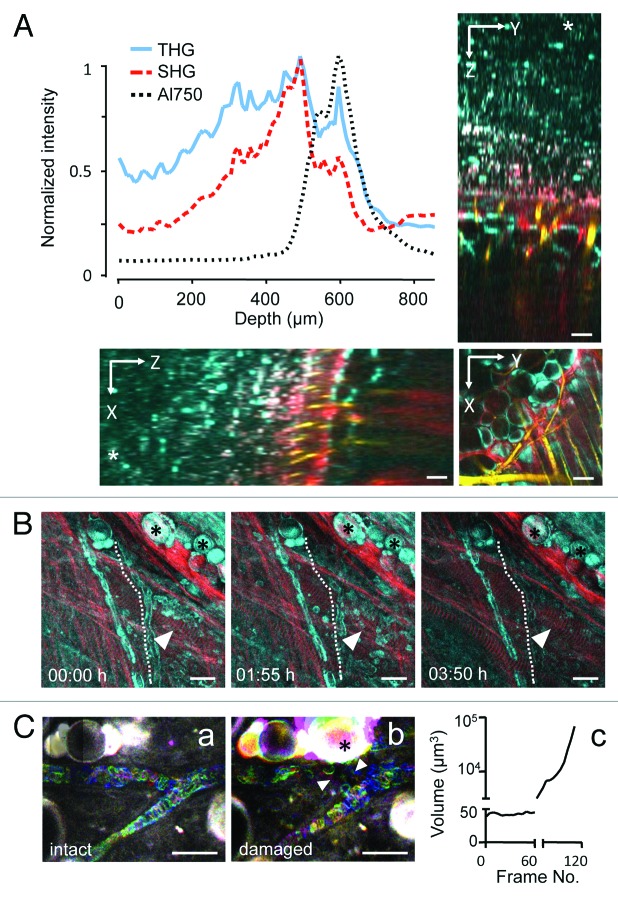
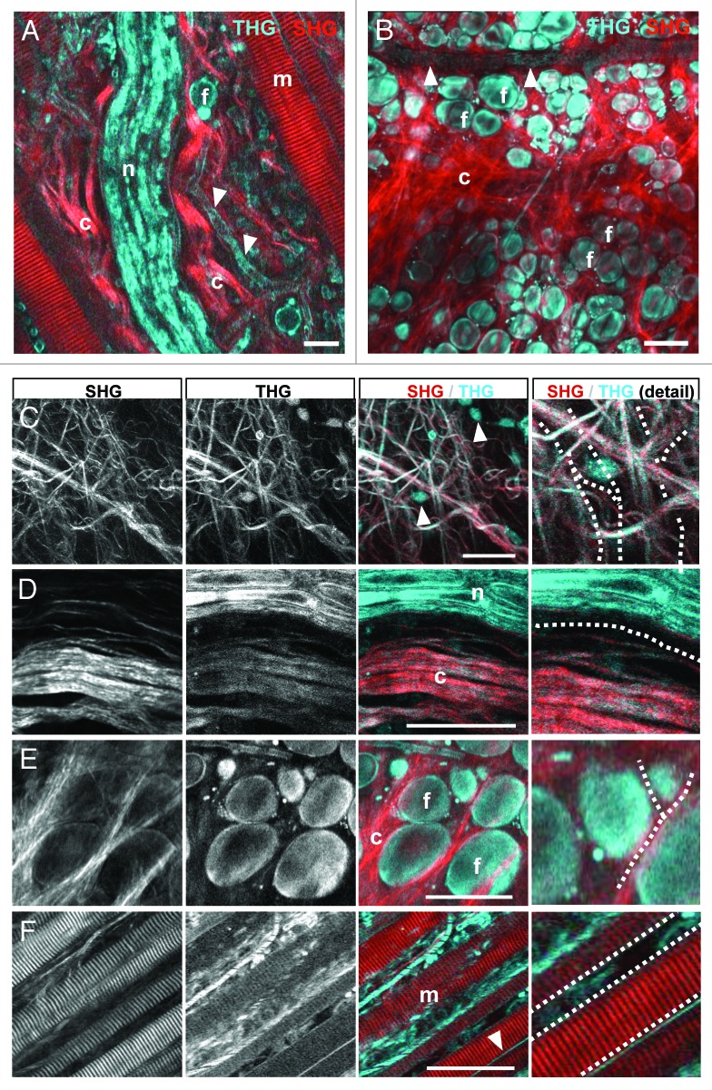
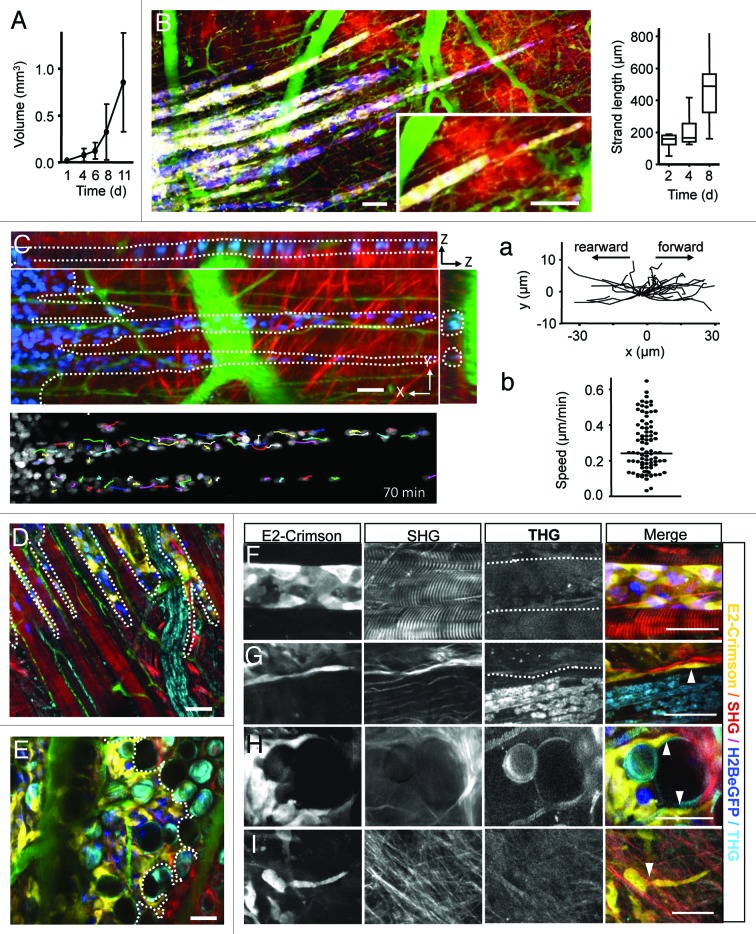
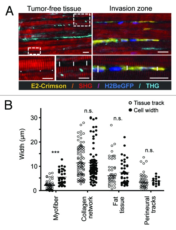
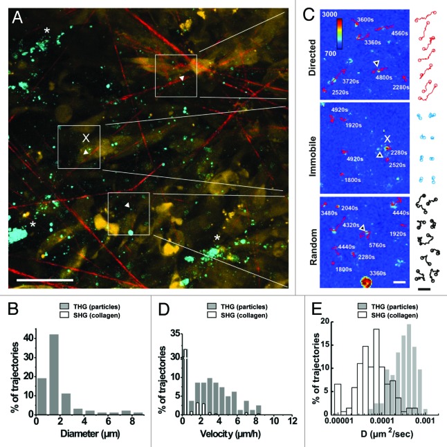
Cancer cell invasion is an adaptive process based on cell-intrinsic properties to migrate individually or collectively, and their adaptation to encountered tissue structure acting as barrier or providing guidance. Whereas molecular and physical mechanisms of cancer invasion are well-studied in 3D in vitro models, their topographic relevance, classification and validation toward interstitial tissue organization in vivo remain incomplete. Using combined intravital third and second harmonic generation (THG, SHG), and three-channel fluorescence microscopy in live tumors, we here map B16F10 melanoma invasion into the dermis with up to 600 µm penetration depth and reconstruct both invasion mode and tissue tracks to establish invasion routes and outcome. B16F10 cells preferentially develop adaptive invasion patterns along preformed tracks of complex, multi-interface topography, combining single-cell and collective migration modes, without immediate anatomic tissue remodeling or destruction. The data suggest that the dimensionality (1D, 2D, 3D) of tissue interfaces determines the microanatomy exploited by invading tumor cells, emphasizing non-destructive migration along microchannels coupled to contact guidance as key invasion mechanisms. THG imaging further detected the presence and interstitial dynamics of tumor-associated microparticles with submicron resolution, revealing tumor-imposed conditioning of the microenvironment. These topographic findings establish combined THG, SHG and fluorescence microscopy in intravital tumor biology and provide a template for rational in vitro model development and context-dependent molecular classification of invasion modes and routes.
癌细胞侵袭是一个适应性过程,基于细胞内在特性进行单独或集体迁移,并使其适应作为屏障或提供引导的所遇到的组织结构。虽然在三维体外模型中对癌症侵袭的分子和物理机制进行了充分研究,但它们在体内对间质组织组织的地形相关性、分类和验证仍不完整。利用活体肿瘤中的活体三次谐波和二次谐波产生(THG、SHG)以及三通道荧光显微镜技术,我们在此绘制了B16F10黑色素瘤侵入真皮的情况,穿透深度可达600微米,并重建了侵袭模式和组织轨迹,以确定侵袭途径和结果。B16F10细胞优先沿着复杂的多界面地形的预先形成的轨迹发展适应性侵袭模式,结合单细胞和集体迁移模式,而不会立即进行解剖组织重塑或破坏。数据表明,组织界面的维度(一维、二维、三维)决定了侵袭肿瘤细胞所利用的微观解剖结构,强调沿着与接触引导相关的微通道进行非破坏性迁移是关键的侵袭机制。THG成像进一步以亚微米分辨率检测到肿瘤相关微粒的存在和间质动态,揭示了肿瘤对微环境的影响。这些地形学发现确立了活体肿瘤生物学中THG、SHG和荧光显微镜的联合应用,并为合理的体外模型开发以及侵袭模式和途径的上下文相关分子分类提供了模板。