Sternecker Katharina, Geist Juergen, Beggel Sebastian, Dietz-Laursonn Kristin, de la Fuente Matias, Frank Hans-Georg, Furia John P, Milz Stefan, Schmitz Christoph
Extracorporeal Shock Wave Research Unit, Chair of Neuroanatomy, Institute of Anatomy, Faculty of Medicine, LMU Munich, 80336 Munich, Germany.
Aquatic System Biology Unit, Department of Ecology and Ecosystem Management, Technical University of Munich, 85354 Freising, Germany.
Biol Open. 2018 Jul 2;7(7):bio033258. doi: 10.1242/bio.033258.
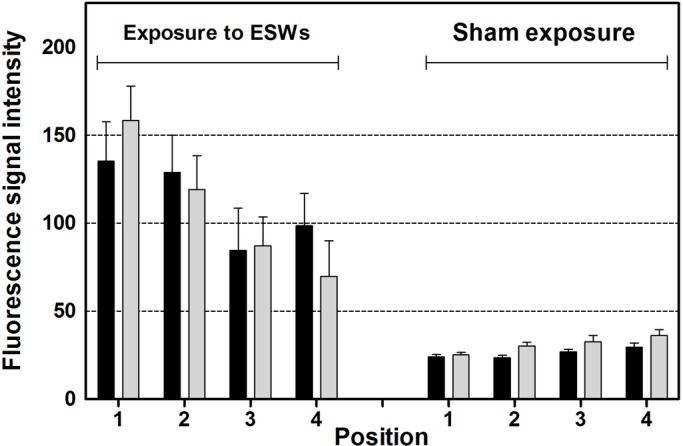
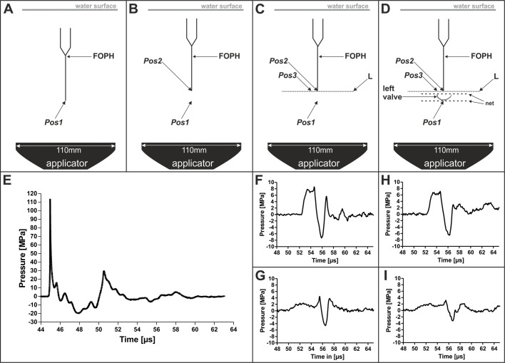
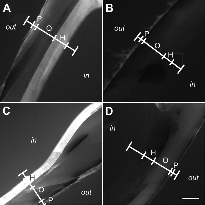
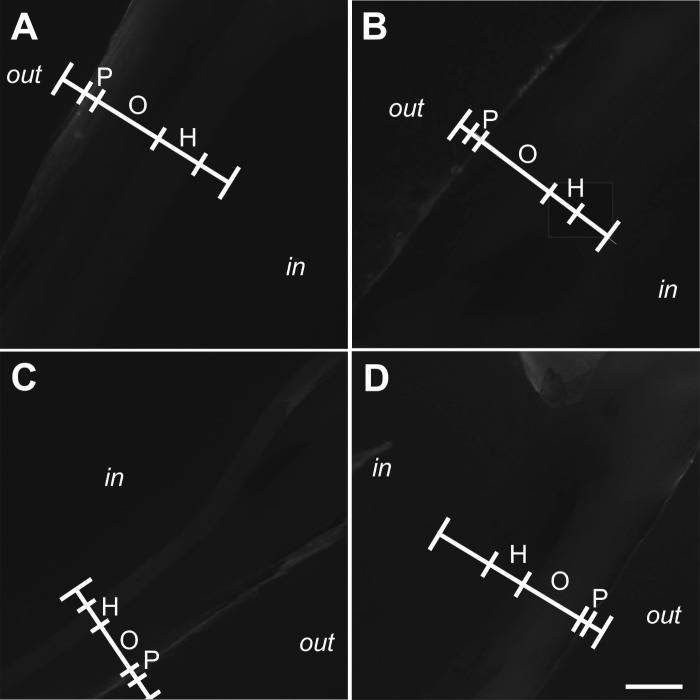
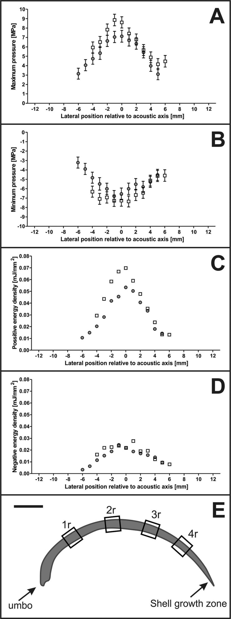
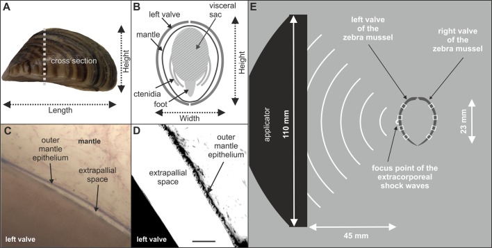
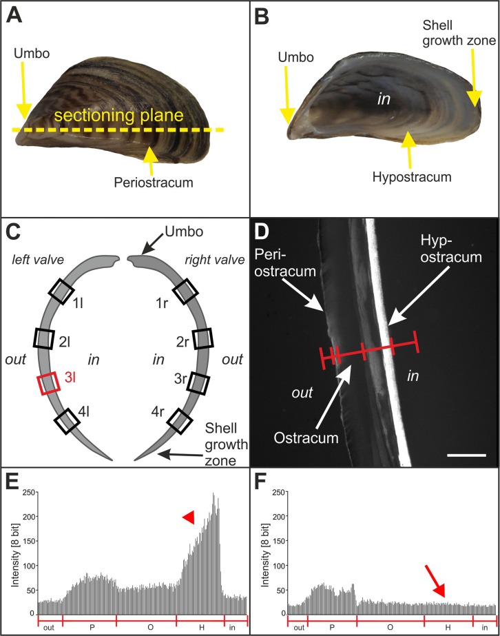
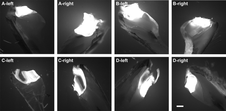
The success rate of extracorporeal shock wave therapy (ESWT) for fracture nonunions in human medicine (i.e. radiographic union at 6 months after ESWT) is only approximately 75%. Detailed knowledge regarding the underlying mechanisms that induce bio-calcification after ESWT is limited. We analyzed the biological response within mineralized tissue of a new invertebrate model organism, the zebra mussel , after exposure with extracorporeal shock waves (ESWs). Mussels were exposed to ESWs with positive energy density of 0.4 mJ/mm (A) or were sham exposed (B). Detection of newly calcified tissue was performed by exposing the mussels to fluorescent markers. Two weeks later, the A-mussels showed a higher mean fluorescence signal intensity within the shell zone than the B-mussels (<0.05). Acoustic measurements revealed that the increased mean fluorescence signal intensity within the shell of the A-mussels was independent of the size and position of the focal point of the ESWs. These data demonstrate that induction of bio-calcification after ESWT may not be restricted to the region of direct energy transfer of ESWs into calcified tissue. The results of the present study are of relevance for better understanding of the molecular and cellular mechanisms that induce formation of new mineralized tissue after ESWT.
在人类医学中,体外冲击波疗法(ESWT)治疗骨折不愈合的成功率(即ESWT后6个月时影像学显示骨折愈合)仅约为75%。关于ESWT后诱导生物钙化的潜在机制的详细知识有限。我们分析了一种新的无脊椎动物模型生物斑马贻贝在暴露于体外冲击波(ESW)后矿化组织内的生物学反应。将贻贝暴露于正能量密度为0.4 mJ/mm的ESW下(A组)或进行假暴露(B组)。通过将贻贝暴露于荧光标记物来检测新钙化组织。两周后,A组贻贝壳区域内的平均荧光信号强度高于B组贻贝(<0.05)。声学测量表明,A组贻贝壳内平均荧光信号强度的增加与ESW焦点的大小和位置无关。这些数据表明,ESWT后生物钙化的诱导可能不限于ESW直接能量转移到钙化组织的区域。本研究结果对于更好地理解ESWT后诱导新矿化组织形成的分子和细胞机制具有重要意义。