Ji Lanxin, Pearlson Godfrey D, Hawkins Keith A, Steffens David C, Guo Hua, Wang Lihong
Center for Biomedical Imaging Research, Department of Biomedical Engineering, Tsinghua University, Beijing, China.
Olin Neuropsychiatry Research Center, Institute of Living, Hartford Hospital, Hartford, CT, United States.
Front Aging Neurosci. 2018 Mar 19;10:71. doi: 10.3389/fnagi.2018.00071. eCollection 2018.
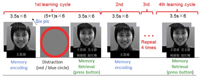
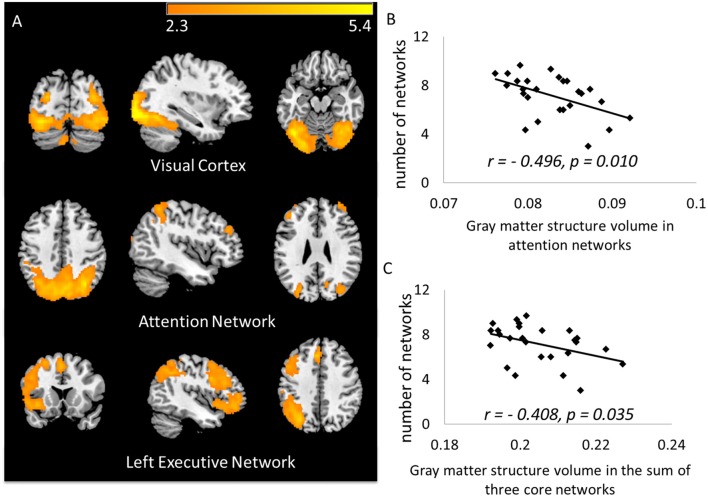
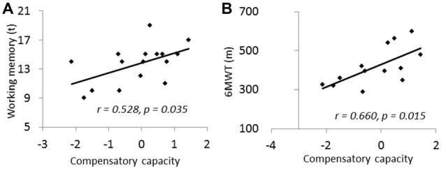
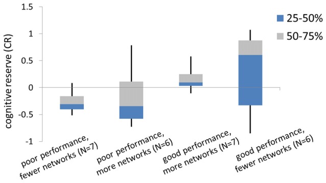
Neuroimaging studies suggest that older adults may compensate for declines in brain function and cognition through reorganization of neural resources. A limitation of prior research is reliance on between-group comparisons of neural activation (e.g., younger vs. older), which cannot be used to assess compensatory ability quantitatively. It is also unclear about the relationship between compensatory ability with cognitive function or how other factors such as physical exercise modulates compensatory ability. Here, we proposed a data-driven method to semi-quantitatively measure neural compensation under a challenging cognitive task, and we then explored connections between neural compensation to cognitive engagement and cognitive reserve (CR). Functional and structural magnetic resonance imaging scans were acquired for 26 healthy older adults during a face-name memory task. Spatial independent component analysis (ICA) identified visual, attentional and left executive as core networks. Results show that the smaller the volumes of the gray matter (GM) structures within core networks, the more networks were needed to conduct the task ( = -0.408, = 0.035). Therefore, the number of task-activated networks controlling for the GM volume within core networks was defined as a measure of neural compensatory ability. We found that compensatory ability correlated with working memory performance ( = 0.528, = 0.035). Among subjects with good memory task performance, those with higher CR used fewer networks than subjects with lower CR. Among poor-performance subjects, those using more networks had higher CR. Our results indicated that using a high cognitive-demanding task to measure the number of activated neural networks could be a useful and sensitive measure of neural compensation in older adults.
神经影像学研究表明,老年人可能通过神经资源的重新组织来补偿大脑功能和认知能力的下降。先前研究的一个局限性在于依赖神经激活的组间比较(例如,年轻人与老年人),而这种比较无法用于定量评估补偿能力。此外,补偿能力与认知功能之间的关系,以及诸如体育锻炼等其他因素如何调节补偿能力也尚不清楚。在此,我们提出了一种数据驱动的方法,用于在具有挑战性的认知任务下对神经补偿进行半定量测量,然后我们探讨了神经补偿与认知参与和认知储备(CR)之间的联系。在一项面孔-名字记忆任务中,对26名健康老年人进行了功能和结构磁共振成像扫描。空间独立成分分析(ICA)确定视觉、注意力和左执行网络为核心网络。结果表明,核心网络内灰质(GM)结构的体积越小,完成任务所需的网络就越多(r = -0.408,p = 0.035)。因此,控制核心网络内GM体积的任务激活网络数量被定义为神经补偿能力的一种度量。我们发现补偿能力与工作记忆表现相关(r = 0.528,p = 0.035)。在记忆任务表现良好的受试者中,CR较高的受试者比CR较低的受试者使用的网络更少。在表现较差的受试者中,使用更多网络的受试者CR较高。我们的结果表明,使用高认知需求任务来测量激活神经网络的数量可能是一种有用且敏感的老年人神经补偿测量方法。