Baltieri Natali, Guizoni Daniele M, Victorio Jamaira A, Davel Ana P
Department of Structural and Functional Biology, Institute of Biology, University of Campinas, Campinas, Brazil.
Front Physiol. 2018 Mar 19;9:229. doi: 10.3389/fphys.2018.00229. eCollection 2018.
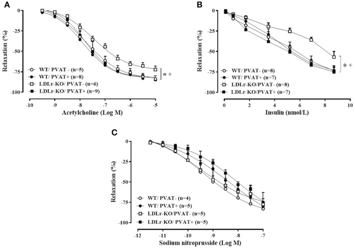
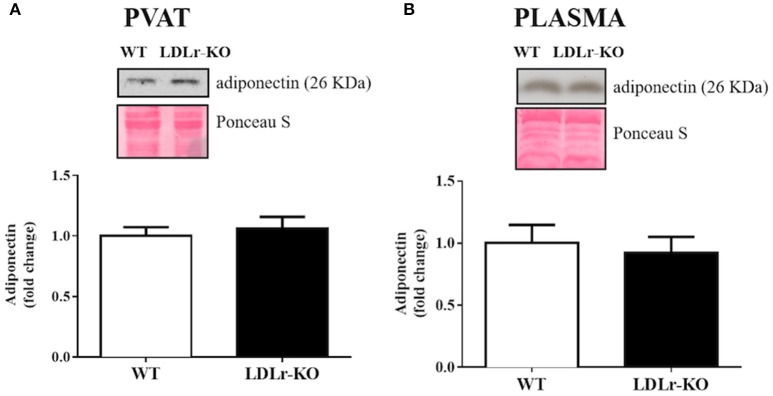
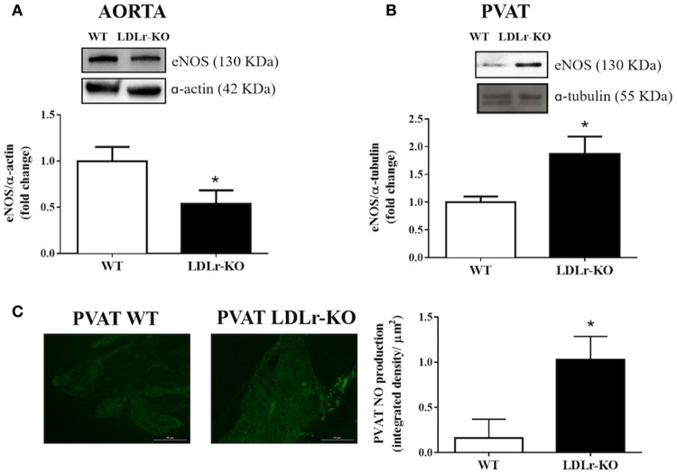
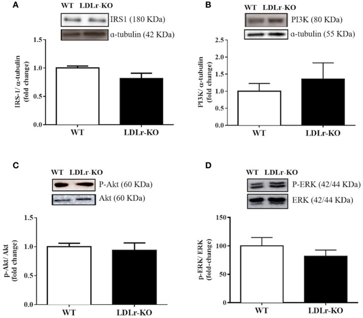
Endothelial dysfunction plays a pivotal role in the initiation of atherosclerosis. Vascular insulin resistance might contribute to a reduction in endothelial nitric oxide (NO) production, leading to impaired endothelium-dependent relaxation in cardiometabolic diseases. Because perivascular adipose tissue (PVAT) controls endothelial function and NO bioavailability, we hypothesized a role for this fat deposit in the vascular complications associated with the initial stages of atherosclerosis. Therefore, we investigated the potential involvement of PVAT in the early endothelial dysfunction in hypercholesterolemic LDL receptor knockout mice (LDLr-KO). Thoracic aortas with and without PVAT were isolated from 4-month-old C57BL/6J (WT) and LDLr-KO mice. The contribution of PVAT to relaxation responses to acetylcholine, insulin, and sodium nitroprusside was investigated. Western blotting was used to examine endothelial NO synthase (eNOS) and adiponectin expression, as well the insulin signaling pathway in aortic PVAT. PVAT-free aortas of LDLr-KO mice exhibited impaired acetylcholine- and insulin-induced relaxation compared with those of WT mice. Both vasodilatory responses were restored by the presence of PVAT in LDLr-KO mice, associated with enhanced acetylcholine-induced NO levels. PVAT did not change vasodilatory responses to acetylcholine and insulin in WT mice, while vascular relaxation evoked by the NO donor sodium nitroprusside was not modified by either genotype or PVAT. The expression of insulin receptor substrate-1 (IRS-1), phosphatidylinositol 3-kinase (PI3K), AKT, ERK1/2, phosphorylation of AKT (Ser473) and ERK1/2 (Thr202/Tyr204), and adiponectin was similar in the PVAT of WT and LDLr-KO mice, suggesting no changes in PVAT insulin signaling. However, eNOS expression was enhanced in the PVAT of LDLr-KO mice, while eNOS expression was less abundant in PVAT-free aortas. These results suggest that elevated eNOS-derived NO production in aortic PVAT might be a compensatory mechanism for the endothelial dysfunction and impaired vasodilator action of insulin in hypercholesterolemic LDLr-deficient mice. This protective effect may limit the progression of atherosclerosis in genetic hypercholesterolemia in the absence of an atherogenic diet.
内皮功能障碍在动脉粥样硬化的起始过程中起关键作用。血管胰岛素抵抗可能导致内皮一氧化氮(NO)生成减少,进而在心脏代谢疾病中引起内皮依赖性舒张功能受损。由于血管周围脂肪组织(PVAT)控制内皮功能和NO生物利用度,我们推测这种脂肪沉积在动脉粥样硬化初始阶段相关的血管并发症中发挥作用。因此,我们研究了PVAT在高胆固醇血症低密度脂蛋白受体敲除小鼠(LDLr-KO)早期内皮功能障碍中的潜在作用。从4个月大的C57BL/6J(野生型,WT)和LDLr-KO小鼠中分离出带或不带PVAT的胸主动脉。研究了PVAT对乙酰胆碱、胰岛素和硝普钠舒张反应的影响。采用蛋白质免疫印迹法检测主动脉PVAT中内皮型一氧化氮合酶(eNOS)和脂联素的表达以及胰岛素信号通路。与WT小鼠相比,LDLr-KO小鼠去除PVAT的主动脉对乙酰胆碱和胰岛素诱导的舒张功能受损。LDLr-KO小鼠中PVAT的存在恢复了这两种血管舒张反应,同时乙酰胆碱诱导的NO水平升高。PVAT对WT小鼠对乙酰胆碱和胰岛素的血管舒张反应没有影响,而NO供体硝普钠引起的血管舒张不受基因型或PVAT的影响。WT和LDLr-KO小鼠的PVAT中胰岛素受体底物-1(IRS-1)、磷脂酰肌醇3激酶(PI3K)、AKT、ERK1/2、AKT(Ser473)和ERK1/2(Thr202/Tyr204)的磷酸化以及脂联素的表达相似,表明PVAT胰岛素信号没有变化。然而,LDLr-KO小鼠的PVAT中eNOS表达增强,而去除PVAT的主动脉中eNOS表达较少。这些结果表明,主动脉PVAT中eNOS衍生的NO生成增加可能是高胆固醇血症LDLr缺陷小鼠内皮功能障碍和胰岛素血管舒张作用受损的一种代偿机制。在没有致动脉粥样硬化饮食的情况下,这种保护作用可能会限制遗传性高胆固醇血症中动脉粥样硬化的进展。