Ma Dongjie, Li Shanqing, Cui Yushang, Li Li, Liu Hongsheng, Chen Yeye, Zhou Xiaoyun
Department of Thoracic Surgery, Peking Union Medical College Hospital, Peking Union Medical College and Chinese Academy of Medical Sciences, Beijing 100730, P.R. China.
Oncol Lett. 2018 May;15(5):6211-6216. doi: 10.3892/ol.2018.8086. Epub 2018 Feb 20.
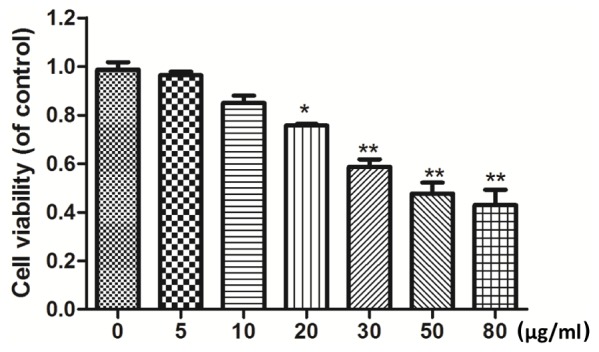
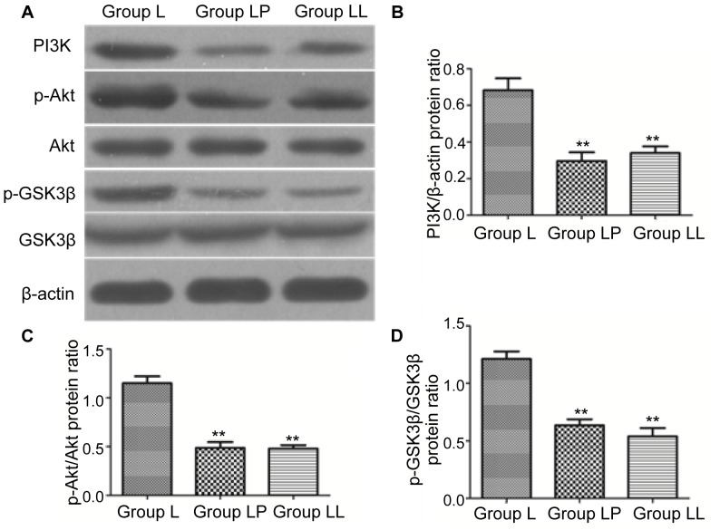
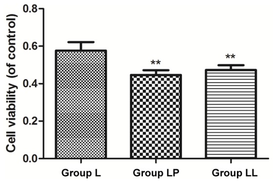
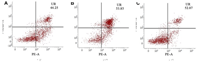
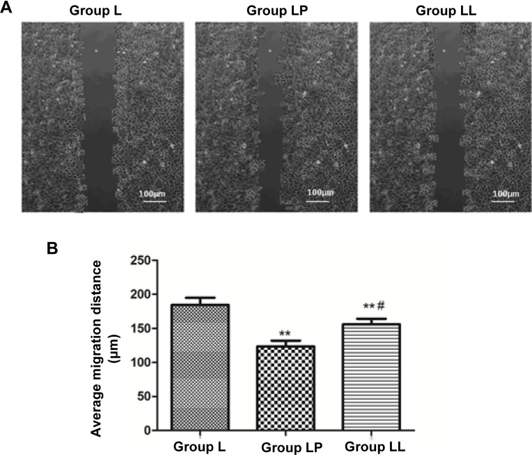
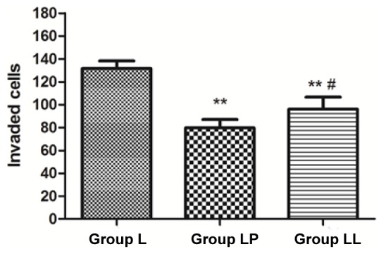
The effect of paclitaxel combined with lobaplatin on the sensitivity of lung cancer cell line NCI-H446 through influencing the phosphatidylinositol 3-kinase (PI3K)/Akt pathway was investigated. The sensitivity of lobaplatin to NCI-H446 and the effect of paclitaxel and PI3K inhibitor LY294002 combined with lobaplatin on the sensitivity to NCI-H446 were detected via methyl thiazolyltetrazolium (MTT) assay. The effect of paclitaxel combined with lobaplatin on cell apoptosis was detected using flow cytometry, the effect of paclitaxel combined with lobaplatin on the cell migration was detected via cell wound scratch assay, and the effect of paclitaxel combined with lobaplatin on the cell invasion was detected via Transwell assay. Finally, the effect of paclitaxel on PI3K/Akt pathway was detected via western blotting. MTT assay showed that 30 µg/ml lobaplatin could significantly inhibit the growth of NCI-H446 (p<0.01). Lobaplatin group (group L), 2 µg/ml paclitaxel combined with lobaplatin group (group LP) and lobaplatin combined with 10 µmol/ml LY294002 group (group LL) were set up. The cell survival rates in group LP and group LL were significantly lower than that in group L (p<0.01), and the cell survival rate in group LP was similar to that in group LL (p>0.05). Flow cytometry revealed that the cell apoptotic levels in group LP and group LL were obviously higher than that in group L (p<0.01), and there was no statistically significant difference in the cell apoptotic level between group LP and group LL (p>0.05). Cell wound scratch assay showed that the cell migration capacity in group LP was significantly lower than those in group L and group LL (p<0.01, p<0.05), and the cell migration capacity in group LL was lower than that in group L (p<0.05). Besides, Transwell assay revealed that the cell invasion capacity in group LP was obviously lower than those in group L and group LL (p<0.01, p<0.05), and the cell invasion capacity in group LL was lower than that in group L (p<0.01). Finally, western blotting showed that the levels of PI3K, phosphorylated-Akt (p-Akt) and phosphorylated-glycogen synthase kinase 3β (p-GSK3β) in group LP and group LL were significantly lower than those in group L, and the differences were statistically significant (p<0.01). Paclitaxel can significantly increase the sensitivity of lobaplatin to lung cancer cell line NCI-H446. Moreover, paclitaxel can enhance the effect of lobaplatin on lung cancer cells and reduce the drug resistance through inhibiting PI3K/Akt pathway.
研究了紫杉醇联合洛铂通过影响磷脂酰肌醇3-激酶(PI3K)/Akt信号通路对肺癌细胞系NCI-H446敏感性的影响。采用甲基噻唑基四氮唑蓝(MTT)法检测洛铂对NCI-H446的敏感性以及紫杉醇和PI3K抑制剂LY294002联合洛铂对NCI-H446敏感性的影响。采用流式细胞术检测紫杉醇联合洛铂对细胞凋亡的影响,通过细胞划痕实验检测紫杉醇联合洛铂对细胞迁移的影响,通过Transwell实验检测紫杉醇联合洛铂对细胞侵袭的影响。最后,通过蛋白质免疫印迹法检测紫杉醇对PI3K/Akt信号通路的影响。MTT法检测结果显示,30μg/ml洛铂可显著抑制NCI-H446细胞的生长(p<0.01)。设置洛铂组(L组)、2μg/ml紫杉醇联合洛铂组(LP组)和洛铂联合10μmol/ml LY294002组(LL组)。LP组和LL组的细胞存活率显著低于L组(p<0.01),且LP组与LL组的细胞存活率相似(p>0.05)。流式细胞术检测结果显示,LP组和LL组的细胞凋亡水平明显高于L组(p<0.01),且LP组与LL组的细胞凋亡水平差异无统计学意义(p>0.05)。细胞划痕实验结果显示,LP组的细胞迁移能力显著低于L组和LL组(p<0.01,p<0.05),且LL组的细胞迁移能力低于L组(p<0.05)。此外,Transwell实验结果显示,LP组的细胞侵袭能力明显低于L组和LL组(p<0.01,p<0.05),且LL组的细胞侵袭能力低于L组(p<0.01)。最后,蛋白质免疫印迹法检测结果显示,LP组和LL组中PI3K、磷酸化Akt(p-Akt)和磷酸化糖原合酶激酶3β(p-GSK3β)的水平显著低于L组,差异具有统计学意义(p<0.01)。紫杉醇可显著提高洛铂对肺癌细胞系NCI-H446的敏感性。此外,紫杉醇可增强洛铂对肺癌细胞的作用,并通过抑制PI3K/Akt信号通路降低耐药性。