Division of Biochemical Toxicology, HFT-110, National Center for Toxicological Research (NCTR), Food and Drug Administration (FDA), 3900 NCTR Road, Jefferson, AR, 72079, USA.
Division of Bioinformatics and Biostatistics, National Center for Toxicological Research (NCTR)/U.S. FDA, Jefferson, AR, 72079, USA.
Arch Toxicol. 2018 Jun;92(6):1969-1981. doi: 10.1007/s00204-018-2196-x. Epub 2018 Apr 3.
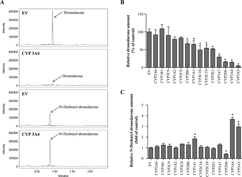
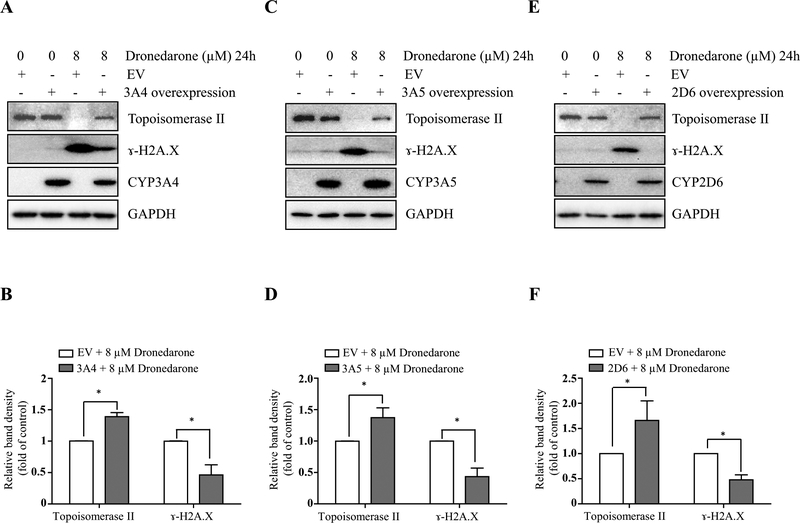
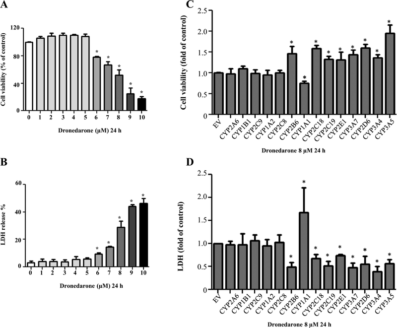
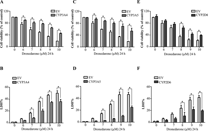
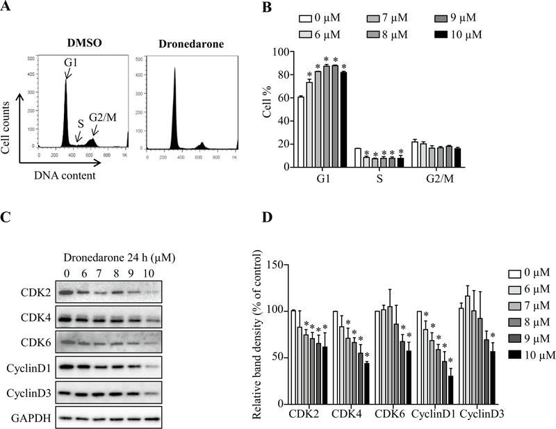
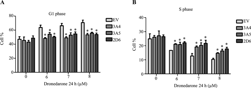
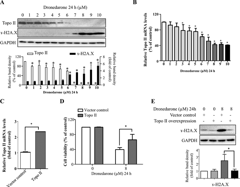
Dronedarone is used to treat patients with cardiac arrhythmias and has been reported to be associated with liver injury. Our previous mechanistic work demonstrated that DNA damage-induced apoptosis contributes to the cytotoxicity of dronedarone. In this study, we examined further the underlying mechanisms and found that after a 24-h treatment of HepG2 cells, dronedarone caused cytotoxicity, G1-phase cell cycle arrest, suppression of topoisomerase II, and DNA damage in a concentration-dependent manner. We also investigated the role of cytochrome P450s (CYPs)-mediated metabolism in the dronedarone-induced toxicity using our previously established HepG2 cell lines expressing individually 14 human CYPs (1A1, 1A2, 1B1, 2A6, 2B6, 2C8, 2C9, 2C18, 2C19, 2D6, 2E1, 3A4, 3A5, and 3A7). We demonstrated that CYP3A4, 3A5, and 2D6 were the major enzymes that metabolize dronedarone, and that CYP3A7, 2E1, 2C19, 2C18, 1A1, and 2B6 also metabolize dronedarone, but to a lesser extent. Our data showed that the cytotoxicity of dronedarone was decreased in CYP3A4-, 3A5-, or 2D6-overexpressing cells compared to the control HepG2 cells, indicating that the parent dronedarone has higher potency than the metabolites to induce cytotoxicity in these cells. In contrast, cytotoxicity was increased in CYP1A1-overexpressing cells, demonstrating that CYP1A1 exerts an opposite effect in dronedarone's toxicity, comparing to CYP3A4, 3A5, or 2D6. We also studied the involvement of topoisomerase II in dronedarone-induced toxicity, and demonstrated that the overexpression of topoisomerase II caused an increase in cell viability and a decrease in γ-H2A.X induction, suggesting that suppression of topoisomerase II may be one of the mechanisms involved in dronedarone-induced liver toxicity.
盐酸多奈哌齐用于治疗心律失常患者,有报道称其与肝损伤有关。我们之前的机制研究表明,DNA 损伤诱导的细胞凋亡导致盐酸多奈哌齐的细胞毒性。在这项研究中,我们进一步研究了潜在的机制,发现经过 24 小时的 HepG2 细胞处理后,盐酸多奈哌齐以浓度依赖性方式引起细胞毒性、G1 期细胞周期停滞、拓扑异构酶 II 抑制和 DNA 损伤。我们还使用我们之前建立的表达个体人细胞色素 P450 (CYP)1A1、1A2、1B1、2A6、2B6、2C8、2C9、2C18、2C19、2D6、2E1、3A4、3A5 和 3A7 的 HepG2 细胞系,研究了 CYP 介导的代谢在盐酸多奈哌齐诱导的毒性中的作用。我们证明 CYP3A4、3A5 和 2D6 是代谢盐酸多奈哌齐的主要酶,而 CYP3A7、2E1、2C19、2C18、1A1 和 2B6 也代谢盐酸多奈哌齐,但程度较低。我们的数据表明,与对照 HepG2 细胞相比,CYP3A4、3A5 或 2D6 过表达细胞中的盐酸多奈哌齐细胞毒性降低,表明母体盐酸多奈哌齐比代谢物对这些细胞的细胞毒性更强。相比之下,CYP1A1 过表达细胞的细胞毒性增加,表明 CYP1A1 在盐酸多奈哌齐的毒性中发挥相反的作用,与 CYP3A4、3A5 或 2D6 相比。我们还研究了拓扑异构酶 II 在盐酸多奈哌齐诱导的毒性中的参与,结果表明拓扑异构酶 II 的过表达导致细胞活力增加和 γ-H2A.X 诱导减少,表明拓扑异构酶 II 的抑制可能是盐酸多奈哌齐诱导的肝毒性的机制之一。